Executive hypothesis
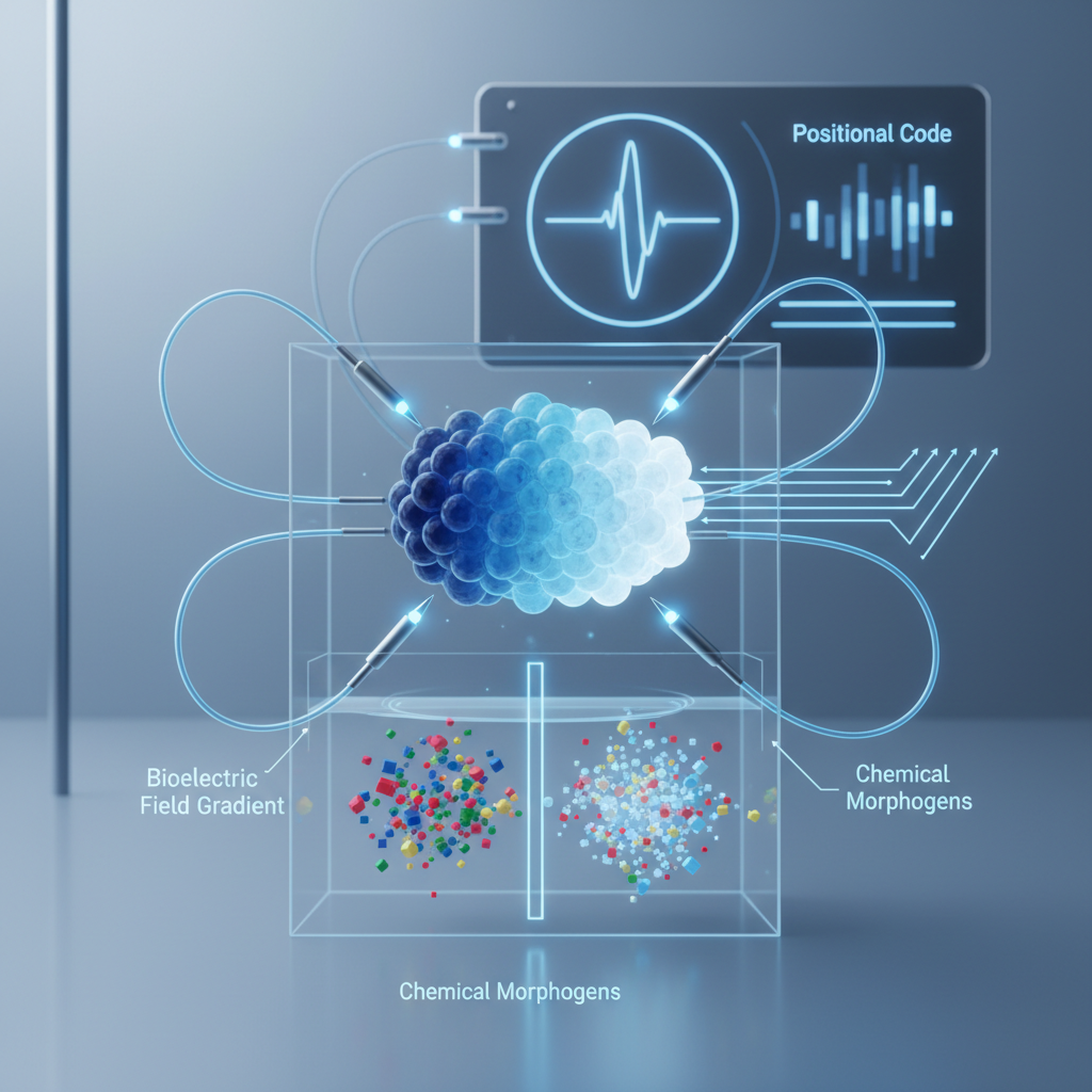
Hypothesis: Spatial gradients of extracellular and transmembrane bioelectric potentials form a robust, information-rich positional code that can specify cell identity and tissue patterning independently of classical chemical morphogens. In many contexts, cells read local vectorial features of the bioelectric field (magnitude, gradient direction, temporal dynamics) to infer their location and execute patterning programs, and artificial manipulation of these fields is sufficient to reprogram positional identity even when morphogen signals are held constant or suppressed.
Why this is plausible
Bioelectric phenomena—membrane potential (Vmem), extracellular potential gradients, ionic fluxes and ephaptic coupling—are pervasive in embryos, regenerating tissues and organoids. Decades of work show that bioelectrical states correlate with developmental events (e.g., axis specification, limb regeneration) and that perturbing ion channels or gap junctions alters morphology. Several properties make bioelectric gradients plausible carriers of positional information:
- Spatial precision: Electric fields vary continuously in space and can form predictable gradient vectors at cellular scale.
- Rapid, long-range propagation: Through gap junction networks and extracellular currents, fields can convey information faster and farther than diffusion-limited morphogens.
- Multiplexing capacity: Voltage magnitude, sign, frequency content and temporal dynamics provide many degrees of freedom for encoding complex coordinates or pattern templates.
- Sensing machinery: Cells express voltage-sensitive proteins (voltage-gated channels, electrosensitive receptors), second-messenger pathways (Ca2+, cAMP) and cytoskeletal responses that transduce electrical cues into gene-expression changes.
Mechanistic sketch
Positional encoding emerges from three interacting components: (1) field generation, (2) local transduction, and (3) interpretation by gene-regulatory networks. Field generation occurs via spatially patterned expression/activity of ion channels and pumps, asymmetric distribution of gap junctions, and tissue geometry producing extracellular potential gradients. These gradients create local transmembrane voltage differences (Vmem) and extracellular electric fields. Cells transduce those electrical cues via voltage-gated Ca2+ channels, electrosensitive membrane enzymes, conformational changes in voltage-sensitive transcriptional regulators, and modulation of charged morphogen transport. The transduced signal modifies local second-messenger concentrations and chromatin states to bias transcription factors (including regionally expressed developmental regulators) toward position-appropriate programs. Importantly, this mechanism allows the same morphogen landscape to be differently interpreted depending on the local electrical context.
Falsifiable predictions
- Imposing a synthetic, stable spatial voltage gradient across a tissue will re-specify positional markers (e.g., Hox or regional patterning genes) in a dose- and vector-dependent manner even when chemical morphogen gradients are flattened.
- Genetic or pharmacological disruption of the ability to sense Vmem (e.g., knockdown of key voltage-gated Ca2+ channels or of a putative voltage-sensitive transcriptional effector) will abrogate positional responses to imposed bioelectric gradients while leaving canonical morphogen pathways intact.
- Cells transplanted between positions in an embryo will adopt host positional identity when host bioelectric gradients are intact; if the host field is abolished but morphogen gradients preserved, transplanted cells will fail to adopt host identity.
- Computational decoding of measured multi-parameter local bioelectric states will predict cell fate outcomes better than local morphogen concentration alone in at least one model system.
Experimental roadmap
Phase 1 — Mapping: use high-resolution voltage reporter imaging (genetically encoded voltage indicators, VSDs) and microelectrode arrays to map spatiotemporal bioelectric landscapes in model systems (Xenopus embryos, planaria, zebrafish embryos, mammalian organoids) during key patterning windows. Correlate electrical features with regional gene expression (in situ hybridization, single-cell RNA-seq).
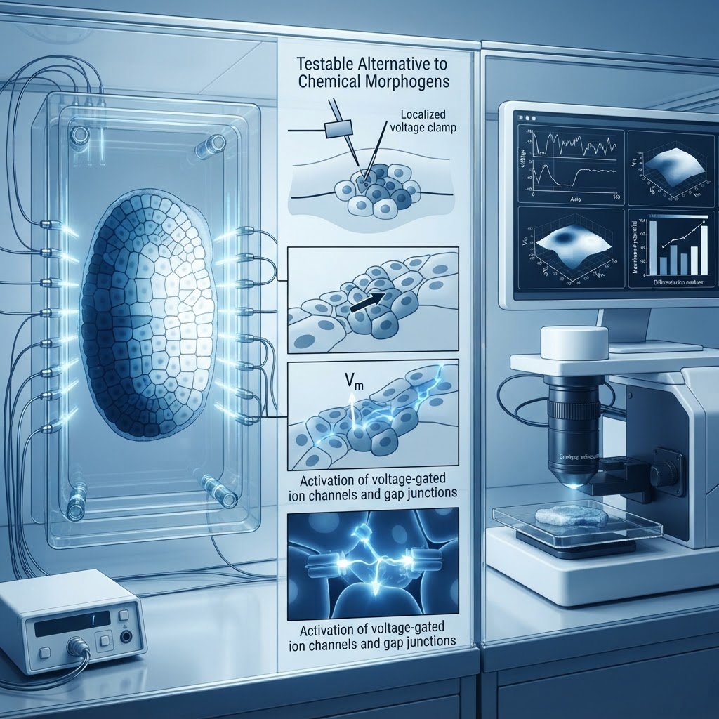
Phase 2 — Causality: impose controlled extracellular potential gradients using microfabricated electrode arrays or optogenetic actuators (e.g., patterned Halorhodopsin/ChR expression) while flattening morphogen distributions via pharmacological inhibitors or uniform morphogen delivery. Assay changes in positional markers, cell behaviors and final morphology.
Phase 3 — Mechanism & decoding: perturb candidate transducers (voltage-gated channels, connexins, electrophysiology-to-nuclear effectors) using CRISPR, dominant negatives and pharmacology; test whether decoding models trained on electrical features predict cell fate. Use closed-loop electrode control to deliver feedback-driven fields that rescue patterning in morphogen-deficient contexts.
Controls and pitfalls
Key controls: sham electrode and light controls, pharmacological specificity controls, maintenance of temperature/pH/ionic strength to exclude nonspecific electrochemical effects, and confirmation that imposed fields do not simply redistribute secreted morphogens (measure morphogen gradients directly). Use genetic mutants lacking major morphogen function to demonstrate independence. Employ rescue experiments where electrical manipulation restores patterning after morphogen pathway disruption.
Potential pitfalls: electrical manipulations can cause cell stress, pH shifts, electrolysis products or ionic concentration changes that indirectly affect development. To mitigate, use low-amplitude, physiologically matched fields; inert electrode materials; and parallel monitoring of ionic composition. Distinguishing direct electrical decoding from secondary effects on morphogen transport requires orthogonal methods: immobilize morphogens (bead tethering), and use impermeant barriers to block convective transport while permitting electrical coupling.
Implications
If validated, this hypothesis establishes a second, parallel positional information system that can operate independently of diffusible morphogens. It would reshape models of tissue patterning, provide a mechanistic basis for many regenerative phenomena, and enable novel bioelectric-based strategies for tissue engineering and repair: programmable electric fields or genetically encoded voltage controllers could specify complex structures without reconstituting every chemical cue. Finally, it links electrophysiology to information theory in development and opens new questions about evolutionary plasticity of patterning codes.