Executive hypothesis
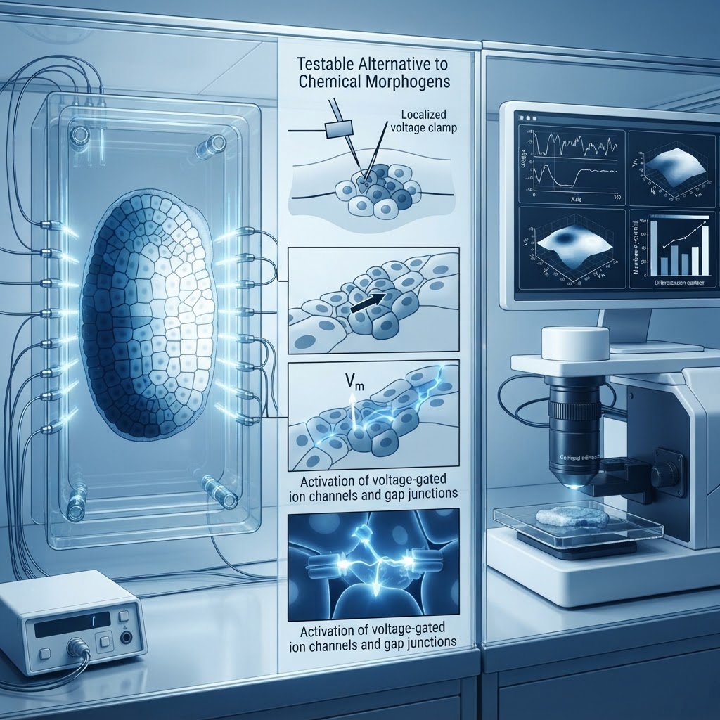
Hypothesis: Spatial gradients of transmembrane potential and resulting bioelectric fields form a robust, information-rich positional code that can specify cell fate and patterning independently of, and in parallel to, classical chemical morphogens. Manipulation of the voltage landscape can reassign positional identities in a manner that is predictable, reproducible, and separable from ligand-receptor signaling.
Why this is plausible
Bioelectric phenomena—membrane potential (Vmem) differences, ionic currents, and field gradients—are ubiquitous across tissues and develop systematically during embryogenesis and regeneration. Several studies demonstrate that Vmem influences proliferation, migration, and gene expression via voltage-sensitive transporters, second-messenger systems, and voltage-dependent transcriptional regulators. Unlike individual morphogen molecules, bioelectric signals are inherently integrative (summating inputs across channels, pumps, and gap junctions), spatially continuous, and can be rapidly redistributed via electrical coupling. These properties allow bioelectric landscapes to encode analog and topological information at scales from single cells to whole organs.
Mechanistic sketch
At the tissue scale, cell membranes establish local Vmem values determined by the complement and activity of ion channels, pumps, and the degree of gap-junctional coupling. Spatial differences in Vmem create electric fields and gradients of ionic flux. Cells read local Vmem through:
- Voltage-gated ion channels altering intracellular ion concentrations and thus signaling cascades (e.g., Ca2+-dependent kinases).
- Voltage-sensitive transporters and pumps that change pH and metabolite gradients affecting epigenetic modifiers.
- Electrogenic modulation of receptor trafficking and mechanotransduction that influence ligand sensitivity.
Information decoding occurs when Vmem-dependent pathways regulate transcription factors that specify positional identity (e.g., Hox genes, regional growth factors). Because gap junctions allow direct electrochemical coupling, a coherent field can propagate positional information over multiple cell diameters without requiring diffusion of a specific morphogen molecule.
Falsifiable predictions
- Establishing an artificial Vmem gradient across a naïve tissue should shift expression domains of positional markers predictably according to the imposed polarity and magnitude, even when classical morphogen gradients are held constant.
- Disrupting gap junctional coupling while preserving local morphogen concentrations will abolish long-range positional coherence if bioelectric fields are primary; conversely, if chemical morphogens are primary, disruption will have limited effect on patterning determined by concentration gradients.
- Optogenetic control of specific ion channels to create temporally patterned Vmem signals should reprogram cell fate decisions in vivo without altering ligand levels, producing reproducible phenotypic changes (e.g., ectopic limb or organ fields in regenerative contexts).
- If bioelectric coding is independent, transcriptomic signatures downstream of imposed Vmem changes will include unique Vmem-responsive gene modules distinct from canonical morphogen-responsive genes.
Experimental roadmap
Model systems: Xenopus laevis embryos, planarian regeneration, zebrafish fin/axis development—systems with demonstrated bioelectric influences.
- Phase 1 — Mapping: High-resolution mapping of Vmem across developmental time using voltage-sensitive dyes and genetically encoded voltage indicators (GEVIs). Correlate Vmem maps with expression domains of positional markers (in situ hybridization, reporter lines).
- Phase 2 — Perturbation: Create defined Vmem gradients using patterned optogenetic actuators (light-gated ion channels/pumps) and microelectrode arrays. Maintain morphogen gradients constant via localized ligand-soaked beads or genetic manipulation. Assess shifts in fate marker domains and tissue morphology.
- Phase 3 — Dissection of pathway: Use single-cell RNA-seq following Vmem manipulation to identify downstream effectors; employ pharmacological blockers and CRISPR knockouts for candidate voltage-transduced mediators (e.g., Ca2+ channels, histone modifiers).
- Phase 4 — Sufficiency and necessity: Ablate gap junctional coupling (dominant-negative connexins) to test requirement for long-range encoding; rescue with engineered electrical networks to demonstrate sufficiency.
Controls and pitfalls
- Controls: sham light stimulation, ion channel mutants lacking conductance, inert beads for morphogen control, and cell-autonomous reporters to rule out indirect effects (e.g., changes in proliferation causing pattern shifts).
- Pitfalls: Voltage dyes and GEVIs can perturb membranes; optogenetic tools may cause ionic imbalance or off-target pH changes. To mitigate, use multiple independent actuators and validate with complementary pharmacology. Distinguishing direct bioelectric effects from secondary chemical changes requires time-resolved assays and rapid actuation where morphogen synthesis cannot respond on the same timescale.
- Interpretation caveat: Bioelectric and chemical systems are likely entangled; demonstrating independence requires orthogonal manipulations and quantitative modeling to show predictive encoding beyond what morphogen concentrations alone can explain.
Implications
If validated, a bioelectric positional code would expand our conceptual toolkit for developmental biology and regenerative medicine: electrical patterning could be harnessed to reprogram tissues non-invasively, guide regeneration, and correct congenital patterning defects. It would imply that positional information is not exclusively carried by diffusible molecules but by an integrated physico-electrochemical field—opening engineering strategies that manipulate information architecture rather than individual genes or ligands.
Conclusion: Testing whether voltage landscapes encode positional information independently is experimentally tractable and conceptually transformative. The proposed roadmap offers explicit, falsifiable steps to adjudicate the hypothesis and, if true, to convert bioelectricity from an obscure regulator into a practical information channel for morphogenetic engineering.