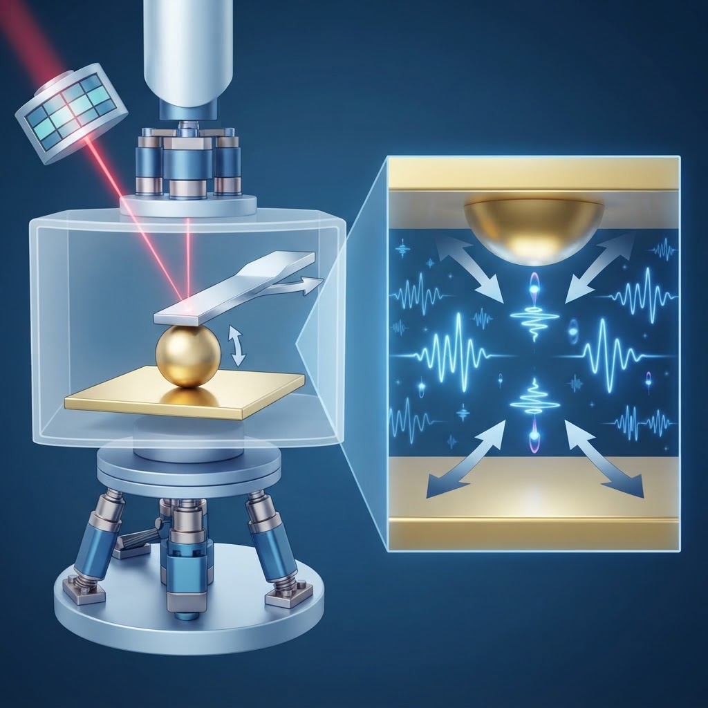
Executive hypothesis
I propose that graded bioelectric fields—established by spatial patterns of membrane potential (Vmem), ion fluxes, and extracellular electric field vectors—constitute an independent and interpretable positional-information code that can specify cellular identity and tissue architecture without relying solely on canonical diffusible chemical morphogens. Manipulating these field gradients will predictably re-specify positional fate and morphogenesis even when morphogen gradients are intact or experimentally held constant.
Why this is plausible
Evidence accumulating from developmental electrophysiology, regeneration studies, and synthetic morphogenesis shows that Vmem gradients correlate with pattern boundaries and that targeted modulation of ion channels alters large-scale patterning. Bioelectric signals are read out by voltage-sensitive transporters, second-messenger systems, and gap-junction networks, and can propagate over cell fields faster and farther than diffusion. The biophysics of extracellular electric fields can bias charged morphogen transport and receptor dynamics, creating a non-chemical dimension of positional cueing. Given cells possess machinery to sense voltage and current (voltage-gated channels, electrosensitive receptors, piezoelectric-associated machinery), a bioelectric gradient is plausibly both producible by embryos and readable by cells as positional information.
Mechanistic sketch
Three nested modules generate and interpret electric positional coordinates:
- Generation: Spatially patterned expression and activity of ion channels, pumps, and gap junctions create stable Vmem gradients across tissues and complementary extracellular electric field vectors. Tissue geometry and extracellular matrix conductivity shape local fields.
- Encoding: Local Vmem differences modulate membrane-localized charge, influence the distribution and kinetics of charged ligands and morphogens, and set thresholds for voltage-dependent transcriptional regulators (e.g., voltage-sensitive phosphatases, calcium channels). The resulting pattern is an analog, graded code (magnitude + vector orientation) that can specify both positional coordinate and polarity.
- Decoding: Cells decode by voltage-dependent ion influx (Ca2+, Na+), electrosensitive transporter activity, polarity shifts of membrane proteins, and gap-junction-mediated patterning of small signaling molecules. This decoding can couple to gene regulatory networks (GRNs) to lock in differentiated states appropriate to inferred position.
Falsifiable predictions
- Creating an artificial extracellular electric field gradient across a tissue, without altering morphogen levels, will elicit spatially predictable changes in cell fate markers and tissue morphology corresponding to the imposed field vector.
- Blocking voltage sensing (e.g., pharmacological blockade of voltage-gated calcium channels and genetic disruption of electrosensitive transducers) will abolish field-imposed positional patterning while leaving morphogen gradients intact, demonstrating causality rather than correlation.
- Recording native Vmem and extracellular field vectors will reveal reproducible, species- and stage-specific spatial patterns that correlate with future morphogenetic axes even before morphogen expression patterns emerge.
- Computational models that combine experimentally measured field gradients with known voltage-dependent decoding kinetics will predict fate boundaries more accurately than models using morphogen concentrations alone.
Experimental roadmap
Stage 1 — Observational mapping: Use voltage-sensitive dyes, extracellular microelectrode arrays, and voltage-reporting genetically encoded indicators to map transmembrane potentials and field vectors across early embryos (zebrafish, Xenopus) and regenerating planaria. Correlate maps with emergent gene-expression boundaries.
Stage 2 — Gain-of-function field imposition: Apply controlled extracellular field gradients with microelectrode arrays or patterned conductive hydrogels while holding morphogen sources constant (localized ligand-soaked beads or inducible expression). Assess fate markers (in situ hybridization, single-cell RNA-seq) and morphogenesis.
Stage 3 — Loss-of-sensing tests: Perturb voltage decoding using pharmacology (VGCC blockers, VSP inhibitors), optogenetic clamps of membrane potential, and CRISPR deletion of candidate electrosensors; test whether imposed fields still instruct patterning.
Stage 4 — Integrative modeling and prediction: Build a biophysical model coupling measured Vmem gradients, extracellular field distribution, charged ligand drift, and intracellular decoding kinetics; validate by prospective prediction of pattern outcomes under novel perturbations.
Controls and pitfalls
Essential controls include: (1) verifying that chemical morphogen concentrations and diffusion are unaltered during field manipulations (via fluorescent ligand quantitation); (2) sham electrical manipulations (no field) to control for device effects; (3) temperature and pH monitoring—electric fields can produce electrochemical side effects; (4) spatially randomized field application to test specificity of vector orientation.
Potential pitfalls: electrolysis and pH shifts at electrodes can cause confounding chemical signals; fields strong enough to perturb cells may induce nonphysiological stress responses; redundancy between bioelectric and chemical cues may mask effects, requiring combinatorial perturbations. Mitigations include AC or pulsed DC to reduce electrolysis, microfluidic isolation of electrodes, careful dosimetry of field strength within physiological ranges, and parallel molecular measurements to detect off-target effects.
Implications
If validated, this hypothesis expands the canonical view of positional information from a purely chemical morphogen-centric paradigm to a multi-modal code that includes bioelectric coordinates. Practical implications span regenerative medicine (programming field gradients to drive limb or organ regrowth), synthetic embryology (designing electric templates to sculpt tissues), and developmental disorder etiology (bioelectric defects as primary causes of patterning defects). Theoretically, an electric coordinate system offers advantages: rapid long-range communication, vectorial information (orientation + magnitude), and integrative coupling to both chemical and mechanical cues—opening a new layer of control over form and organization.