A Signal Hidden in Plain Sight
For most of the twentieth century, the dominant framework of developmental biology was chemical. Morphogen gradients — diffusible molecules like Bicoid, Sonic Hedgehog, and Wnt ligands — were understood to carry the positional information that tells a cell where it is and what to become. This view was extraordinarily productive, yielding Nobel prizes and an entire molecular vocabulary for embryology. Yet it left something unexplained: the speed, robustness, and long-range coordination of certain patterning events that chemical diffusion alone struggles to account for. The answer, accumulating across decades of careful electrophysiology and now accelerated by new optogenetic and chemogenetic tools, points toward a parallel channel of biological instruction — the bioelectric field.
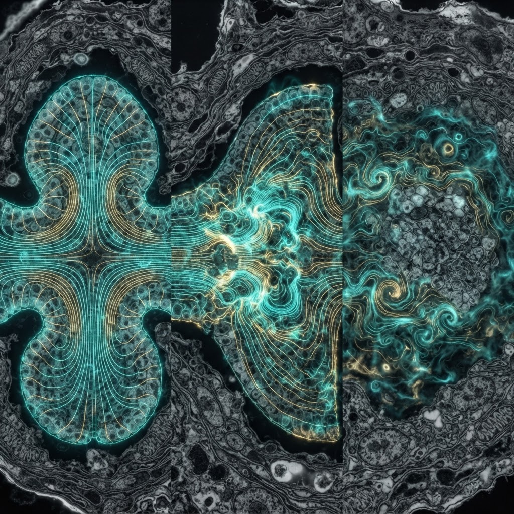
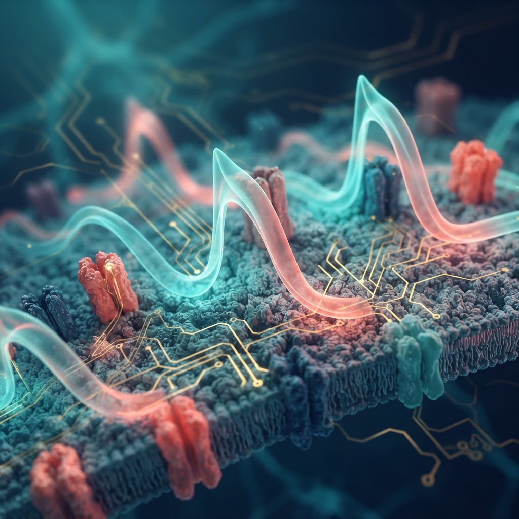
Endogenous bioelectric signals arise from the regulated activity of ion channels, pumps, and gap junctions expressed across cells and tissues. These are not static charges but dynamic, spatially structured voltage landscapes — transmembrane potentials (Vmem) that vary systematically across developing tissues and that change, predictably, when developmental fate changes. The central claim of the bioelectric morphogenesis hypothesis is that this spatial patterning of Vmem is not a downstream readout of chemical decisions but an upstream, causally active regulator of those decisions.
Historical Foundations: From Galvani to Levin
The story begins, awkwardly, with Luigi Galvani's frog legs in the 1780s, which established that living tissue generates and responds to electricity. Harold Saxton Burr at Yale spent the 1930s–1950s mapping what he called the "electrodynamic fields" of organisms, documenting voltage gradients in salamander eggs and developing embryos with primitive but consistent methodology. Burr's work was largely ignored as too vitalistic, too far from the mechanistic mainstream. More rigorous was the work of Lionel Jaffe and Richard Nuccitelli in the 1970s and 1980s, who used the vibrating probe — an electrode that oscillates near a tissue surface to detect ionic currents — to demonstrate that measurable transcellular currents flow through and around developing eggs, regenerating limb stumps, and wound sites. Jaffe's group showed that the vegetal pole of fucoid algal eggs carries a net inward calcium current that predicts the future axis of polarization, and that artificially reversing this current reverses the axis.
The modern synthesis arrived principally through the work of Michael Levin at Tufts University, who, beginning in the early 2000s, moved from descriptive electrophysiology to causal manipulation. Using pharmacological blockers of specific ion channels and, later, optogenetic and chemogenetic actuators, Levin's group demonstrated that misregulating Vmem in Xenopus laevis embryos could produce dramatic and specific anatomical outcomes: ectopic eyes, duplicated body axes, and — most strikingly — the induction of complete, properly patterned heads in planaria at locations defined not by genetics but by electrically imposed positional cues.
Mechanisms: Ion Channels, Gap Junctions, and the Voltage Code
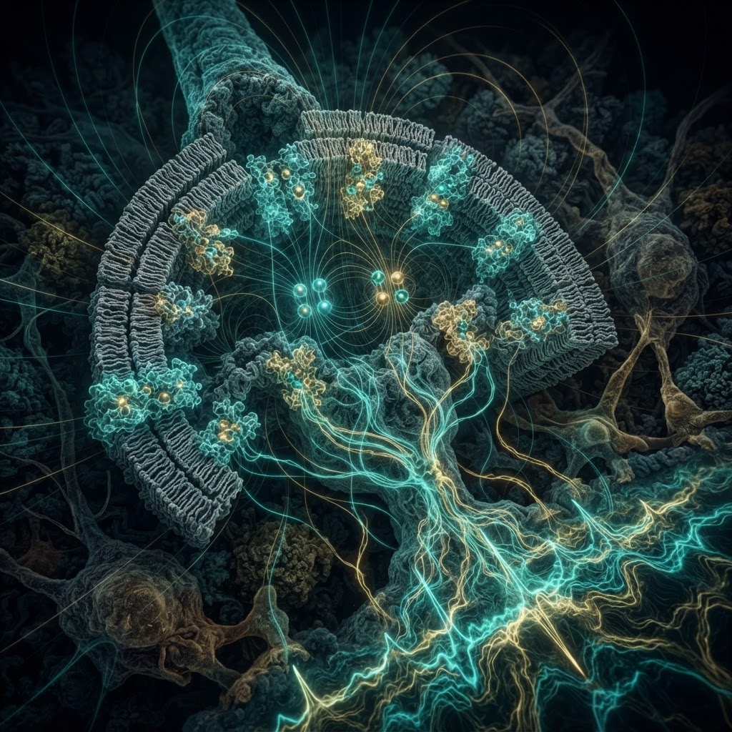
How does a voltage gradient translate into a patterning outcome? The mechanistic picture is now multi-layered. At the cellular level, Vmem directly controls the activity of voltage-gated ion channels, but its morphogenetic effects extend well beyond classical excitable-cell physiology. Key mechanisms include:
- Serotonin transport as a voltage-sensitive relay: The serotonin transporter (SERT) is electrogenic — it moves serotonin with a net charge across the membrane in a voltage-dependent manner. In Xenopus embryos, Levin's group showed that Vmem gradients control intracellular serotonin distribution, which in turn modulates histone modification and gene expression patterns. This provided a concrete molecular path from bioelectric state to transcriptional identity.
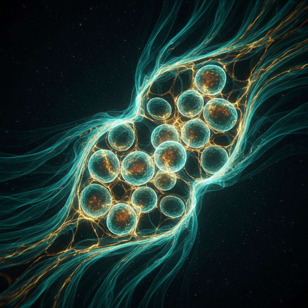
- Gap junctional coupling and syncytial computation: Gap junctions — channels formed by connexin or innexin proteins — connect cells electrically and metabolically. The spatial pattern of gap junctional coupling determines which cells share bioelectric information and on what timescale. Crucially, the gap junction network allows bioelectric states to propagate across tissue lengths far exceeding single-cell dimensions, creating macroscopic voltage domains. Disrupting specific connexins in Xenopus causes laterality defects; in planaria, gap junction manipulation produces double-headed or no-headed worms with high fidelity.
- Vmem-regulated trafficking of morphogenetic receptors: Membrane potential controls the cellular distribution of key signaling receptors, including those for Notch, Wnt, and BMP pathways. A depolarized cell internalizes certain receptors differently than a hyperpolarized one, biasing the transduction of neighboring signals. This creates a bidirectional coupling: bioelectric state modulates chemical signal interpretation, and chemical signals can feed back to alter ion channel expression.
- Electrophoresis of membrane components: In epithelial sheets, laterally directed electric fields — documented at wound edges and along developing axes — can drive the electrophoretic migration of charged membrane proteins, directly sorting receptor landscapes within the plane of the membrane. This mechanism, demonstrated in corneal and skin epithelia, requires no transcription and operates on a timescale of minutes.
Quantitatively, the relevant voltage differences are modest by the standards of action potentials — typically 20–80 mV differences between adjacent tissue regions — but they are sustained over hours to days, and their spatial structure is precisely regulated. In regenerating planarian flatworms, the anterior-pole bioelectric state, characterized by relative hyperpolarization mediated by the H,K-ATPase pump, is established within two hours of amputation and is necessary for correct head-versus-tail identity specification.
Evidence from Model Systems: Planaria, Xenopus, and Beyond
The most dramatic demonstrations of bioelectric morphogenesis come from model systems with strong regenerative capacity. In planaria (Schmidtea mediterranea), pharmacological inhibition of innexin gap junctions during regeneration produces a striking phenotype: worms regenerate a head at both the anterior and posterior wound surfaces, regardless of the original body polarity. This double-headed phenotype persists across subsequent rounds of regeneration even without continued drug treatment — the animals have been stably reprogrammed to a new bioelectric "memory" state. This is remarkable because it suggests that bioelectric patterning can constitute an epigenetic memory orthogonal to DNA sequence.
In Xenopus laevis, modulating the proton pump V-ATPase during early embryogenesis — before most transcription factors critical for eye development are active — is sufficient to induce ectopic eye structures complete with lens, retinal pigment, and correct histological layering. The eyes appear not only on the head but on the trunk and tail, in locations that depend on where the bioelectric manipulation was applied. Comparable results have been obtained for tail regeneration in tadpoles: a brief burst of channelrhodopsin-mediated photostimulation, delivered to the spinal cord stump immediately post-amputation, rescues tail regrowth in an otherwise refractory developmental stage.
In mammalian systems, the evidence is less dramatic but substantial. Human keratinocyte sheets maintain a standing transepithelial potential of 20–40 mV, with the lumen-facing side negative relative to the basal side. This field drives directed cell migration (galvanotaxis) at wound edges with a sensitivity threshold below 10 mV/mm. Disruption of this field by chloride channel blockers significantly impairs wound closure in both in vitro models and rodent skin wounds in vivo.
Competing Hypotheses and Methodological Tensions
The bioelectric morphogenesis field is not without its critics, and the tensions are instructive. A central methodological objection concerns specificity: most pharmacological interventions that alter Vmem (ouabain blocking the Na/K-ATPase, bumetanide blocking NKCC1, ivermectin opening innexin channels) have pleiotropic effects on cell metabolism, osmolarity, and intracellular signaling that are difficult to fully dissociate from voltage effects per se. The development of optogenetic and chemogenetic actuators (channelrhodopsins, DREADDs coupled to ion channels) has substantially improved spatiotemporal specificity, but critics note that even light-gated channels alter calcium dynamics and membrane trafficking independently of voltage changes.
A second tension concerns the hierarchy of signals. Are bioelectric fields truly instructive — capable of overriding genetic programs — or are they permissive modulators that merely bias the probability of chemically specified outcomes? The evidence supports the former in some contexts (the planarian double-head reprogramming that persists through regeneration clearly overrides genetic defaults), but in others the bioelectric effect appears to amplify or sharpen a pre-existing chemical gradient rather than generate de novo pattern. The two roles are not mutually exclusive, and a mature theory will likely accommodate both.
A third debate concerns the physical reality of the "bioelectric code" concept — the idea that specific voltage states encode specific anatomical outcomes in a combinatorial and readable way, analogous to the genetic code. Levin and colleagues have argued for this framework, and computational models based on Ising-type bioelectric state transitions show some predictive power for planarian outcomes. But critics find the analogy imprecise and argue that what is observed is better described as a regulatory network with bioelectric nodes, not a code with discrete symbols.
Open Questions and Future Directions
Several problems sit at the frontier of the field. First, the transduction mechanisms linking Vmem to transcription factor activity remain incompletely mapped in most tissues. The serotonin-histone axis in Xenopus is well characterized, but orthologous pathways in mammalian systems are not yet established. Second, the relationship between bioelectric patterning and the mechanical forces that also shape tissue — cytoskeletal tension, extracellular matrix stiffness, osmotic pressure — is essentially uncharted. Both bioelectric and mechanosensory signals can activate overlapping downstream pathways (YAP/TAZ, Piezo channels, integrins), suggesting deep integration, but the logic of their interaction is unknown.
Third, and perhaps most consequentially for medicine, is the question of bioelectric dysregulation in cancer. Tumor cells are well-documented to be chronically depolarized relative to their normal counterparts, and several groups have reported that inducing hyperpolarization in tumor cells — pharmacologically or optogenetically — reduces proliferation and, in some cases, induces differentiation toward a non-cancerous phenotype. Whether this represents a causal bioelectric vulnerability of cancer or a secondary consequence of metabolic reprogramming remains actively contested.
The tools to resolve these questions are arriving rapidly: genetically encoded voltage indicators (GEVIs) with sufficient sensitivity to image resting membrane potential dynamics in living tissue; light-activated ion pumps and channels with precisely characterized kinetics; single-cell multi-omic profiling capable of linking Vmem state to transcriptional identity at cellular resolution. The coming decade will likely determine whether bioelectric morphogenesis is a parallel layer of developmental control as fundamental as the genetic code — or a sophisticated modulatory system that refines, rather than rewrites, chemical patterning. Either answer would be transformative.