Executive hypothesis
Hypothesis: The historical L-field described by Harold Saxton Burr corresponds to measurable, spatially organized bioelectric potentials arising from patterned distributions of ion channels, pumps, gap-junctional coupling, and extracellular conductivity; these endogenous voltage patterns both report and instruct cell behaviors that drive morphogenesis, regeneration, and early tumorigenesis. Normalization or targeted reconfiguration of these bioelectric patterns is sufficient to redirect aberrant outcomes.
Why this is plausible
Burr's observations (1920s–1950s) that steady voltage patterns precede morphological change anticipated modern discoveries that membrane potential (Vmem) gradients and extracellular electric fields regulate cell proliferation, migration, and gene expression. Contemporary work shows that Vmem gradients are causal in axial patterning and regeneration, and that manipulating ion channels or gap junctions can re-specify anatomy in Xenopus, planaria and organoids (Levin, 2014; McCaig et al., 2005). Thus Burr's phenomenological L-field can be reframed as an emergent property of well-characterized ionic mechanisms.
Mechanistic sketch
The L-field emerges from three interacting components:
- Transmembrane ionic machinery: Spatially patterned expression of ion channels (e.g., Kir, HCN, ENaC), pumps (Na+/K+-ATPase), and exchangers that set local Vmem.
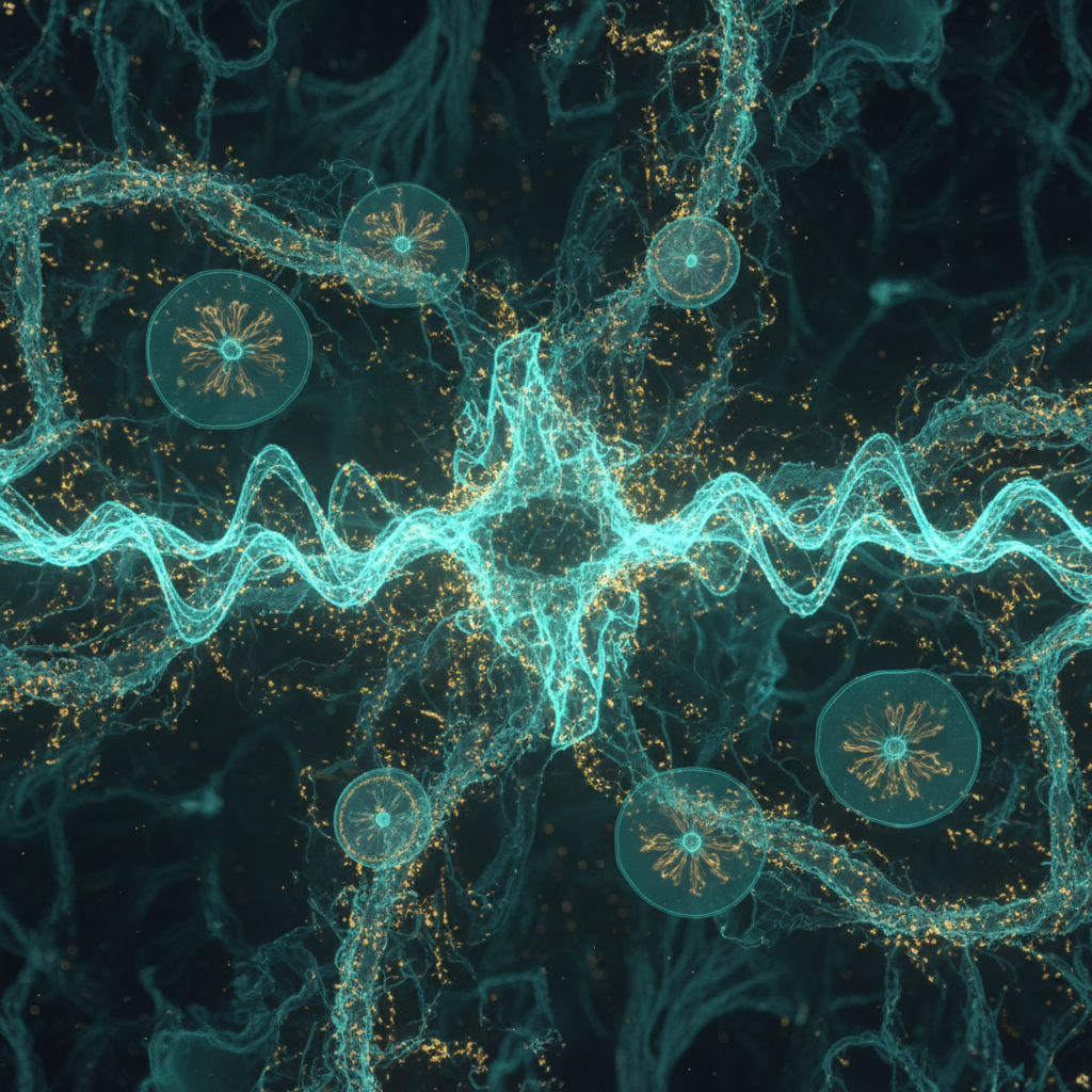
- Intercellular coupling and tissue-scale integration: Electrical coupling via connexin-mediated gap junctions and extracellular ionic conduction create tissue-scale gradients and equipotentials.
- Voltage transduction pathways: Vmem changes modulate intracellular Ca2+ via voltage-gated channels, activate voltage-sensitive enzymes (e.g., voltage-sensitive phosphatases), and bias electrotactic migration and transcriptional programs (Wnt/Notch/TGF-β downstream modulation).
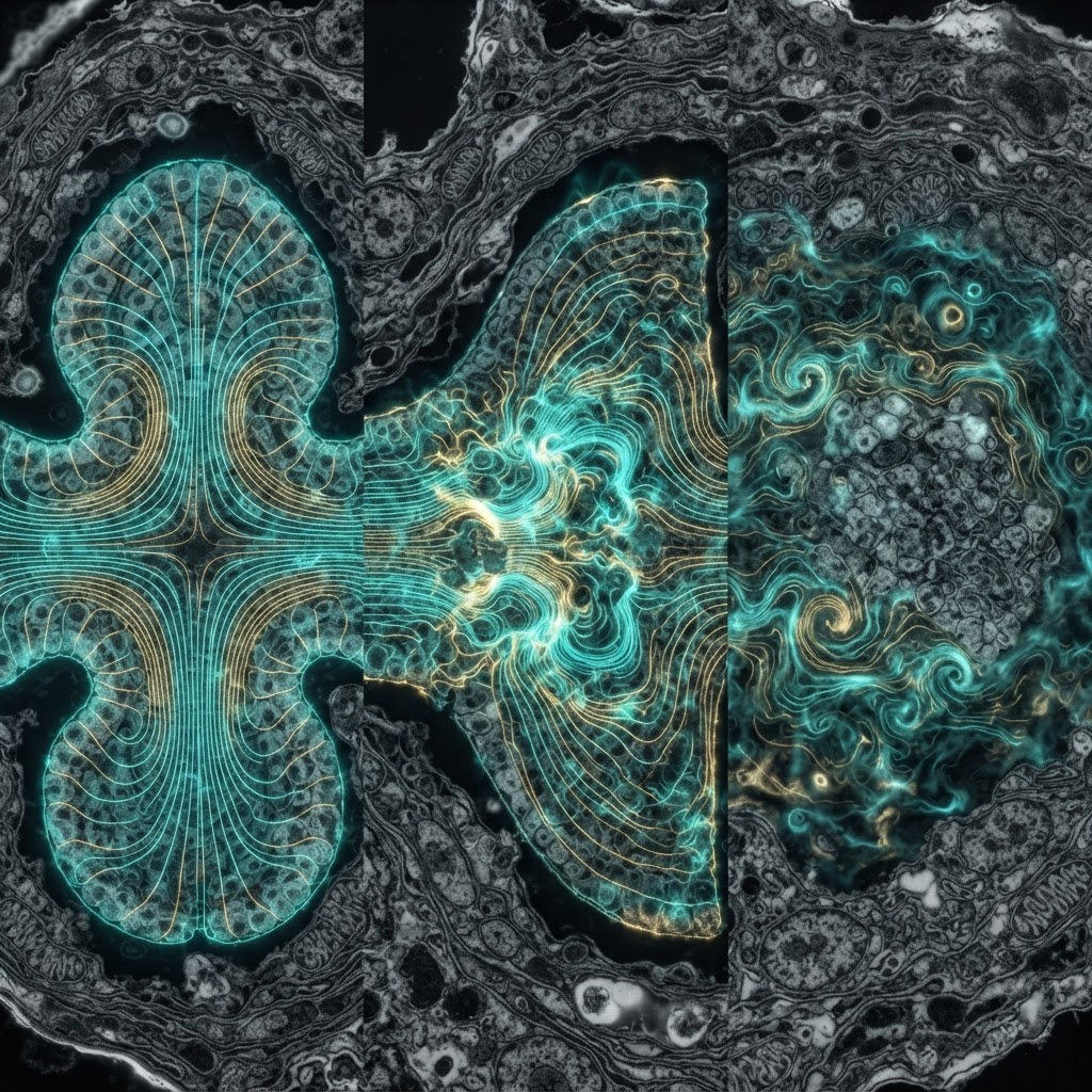
Together these produce stable, reproducible voltage topographies (the L-field) that act as instructive patterning cues; perturbation propagates through the network to alter pattern outcomes.
Falsifiable predictions
- If Burr's L-fields are physiologically meaningful, high-resolution Vmem imaging will reveal spatial patterns that reliably precede specific morphological events during embryogenesis and regeneration.
- Targeted perturbation of predicted ionic contributors (e.g., local expression of Kir2.1 to hyperpolarize a region) will reproducibly alter morphological outcomes in a manner predicted by the preexisting voltage map.
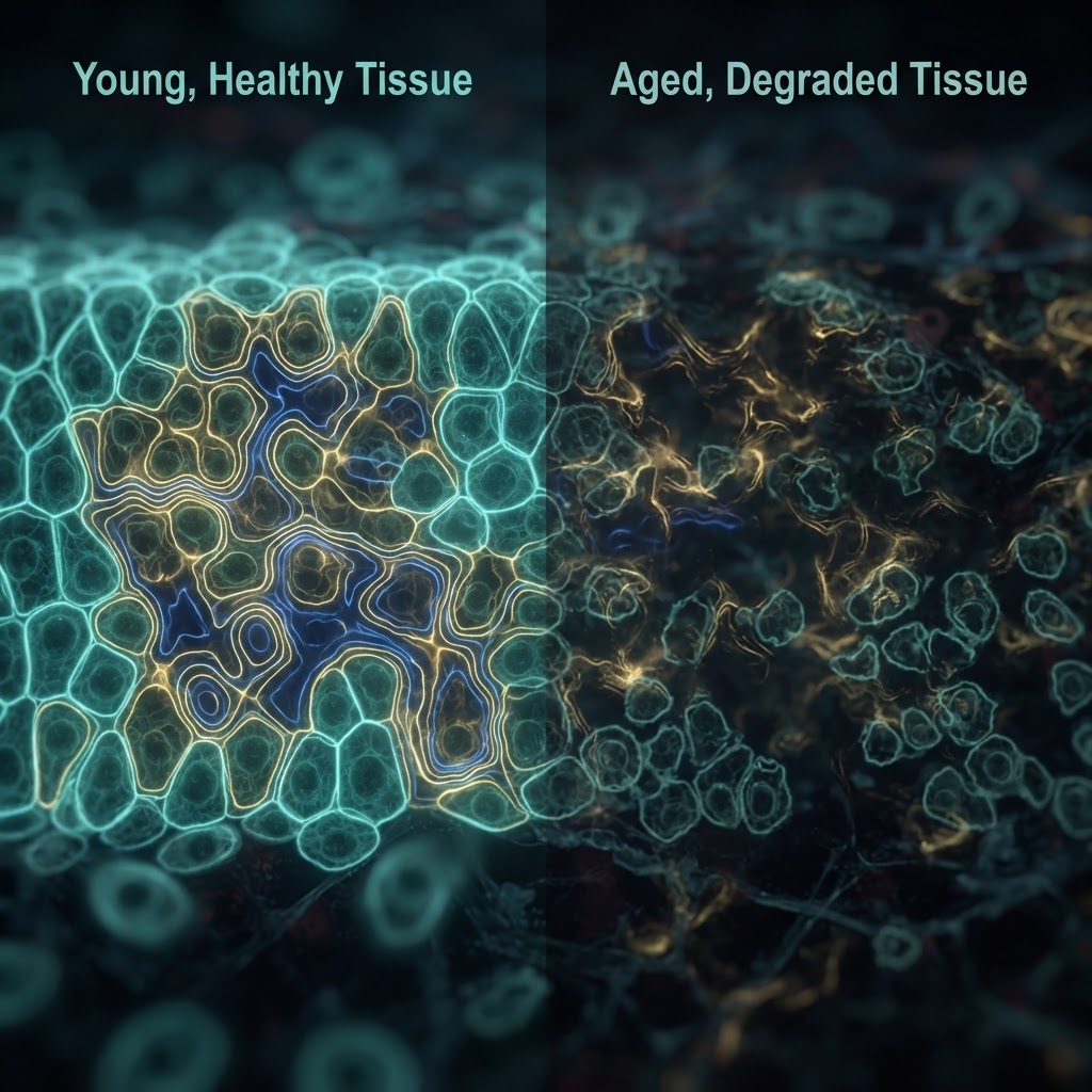
- Distinct, reproducible L-field signatures will distinguish pre-malignant tissue from healthy tissue before histological changes; electrical normalization will reduce tumorigenic behaviors in vitro and in vivo.
- If L-fields are epiphenomenal, then robust alterations of tissue Vmem (within physiological ranges) will not produce consistent changes in patterning, regeneration, or tumor initiation.
Experimental roadmap
Use a layered approach combining descriptive mapping, causal manipulation, and translational tests.
- Phase I — High-resolution mapping: Reproduce Burr's mapping with modern tools. In Xenopus and zebrafish embryos, planaria, and mammalian organoids, map Vmem using genetically encoded voltage indicators (GEVIs, e.g., ASAP-family) and voltage-sensitive dyes across developmental timecourses. Complement with microelectrode arrays (MEAs) and scanning vibrating probe to resolve extracellular fields. Quantify whether voltage topographies precede morphological change.
- Phase II — Causal perturbation: Use targeted optogenetic actuators (e.g., halorhodopsin, channelrhodopsin variants, proton pumps) and ion channel misexpression (Kir2.1, NaV, HCN) driven by spatial promoters or focal electroporation to impose predicted L-field alterations. Measure effects on axis formation, organ primordia, and regeneration outcomes. Monitor downstream signaling (Ca2+ imaging, transcriptional reporters for Wnt/Notch).
- Phase III — Tumor prediction and intervention: In organoid and xenograft models, identify Vmem signatures that precede oncogenic transformation (induced by Ras, Myc). Test whether correcting aberrant Vmem (pharmacologically or optogenetically) reduces proliferation, invasiveness, or tumor-initiation capacity.
- Phase IV — Mechanistic dissection: Use CRISPR to perturb candidate transducers (voltage-sensitive phosphatases, specific connexins) and test for loss of L-field effects. Perform single-cell RNA-seq spatially resolved to link Vmem states to gene programs.
Controls and pitfalls
Key controls: electrode/dye-only shams, non-functional optogenetic mutants, spatially offset perturbations to control for mechanical/thermal effects, and metabolic controls to exclude oxygen/ATP-mediated artifacts. Pitfalls include dye toxicity, motion artifacts, and tissue heating by optogenetics. Distinguish extracellular field artifacts from transmembrane Vmem by combining intracellular recordings, GEVIs, and extracellular probes. Dose–response and temporal causality analyses are essential. Finally, beware confounding by inflammation or necrosis producing spurious potentials.
Implications
If validated, this program will translate Burr's descriptive L-field into a molecularly grounded, predictive bioelectric theory of form: a framework that unifies classical embryology, regenerative biology, and early cancer detection. Practically, it could yield early electrical biomarkers for pathology, non-genetic interventions to guide regenerative outcomes, and new anti-cancer strategies that restore native bioelectric states. It repositions an underappreciated historical insight within a modern, mechanistic rubric — converting Burr's life-field from literary curiosity into experimentally actionable biology.