Overview
The heart is not only an electrochemical pump but also a dynamic electromagnetic source. Magnetocardiography (MCG) provides a window onto the magnetic component of cardiac activity, resolving the geometry and timing of currents that generate the ECG. Recent technical advances have sharpened MCG's ability to probe coherence across cardiac tissue and to ask whether those coherent fields can meaningfully interact with other physiological rhythms — a process often summarized as entrainment. This report synthesizes how MCG measures cardiac magnetic fields, what we mean by coherence patterns and field strength, and the plausible mechanisms and limits of physiological entrainment.
What is magnetocardiography?
Magnetocardiography is the noninvasive measurement of magnetic fields produced by the time-varying currents in the heart. Historically carried out with superconducting quantum interference devices (SQUIDs) in magnetically shielded rooms, the field is now seeing the addition of optically pumped magnetometers (OPMs) that permit closer sensor placement and more flexible montages.
MCG sensors detect the vector magnetic field produced by intracellular and extracellular current loops during depolarization and repolarization. Because magnetic fields are unaffected by the conductivity boundaries that shape electric potentials, MCG can recover source orientations and produce spatially resolved maps (magnetocardiograms) that complement the electrocardiogram (ECG).
Field strength: how large are heart magnetic fields?
Measured cardiac magnetic fields at the chest surface are very small but accessible: typical peak amplitudes are in the picoTesla range. Put numerically, surface fields over the heart are commonly on the order of 10−12 to 10−10 tesla (picotesla, pT), with the precise magnitude depending on sensor distance, patient anatomy, and cardiac moment. These fields decline roughly with distance according to dipolar and more complex source geometry, such that at a meter-scale distance the signal becomes vanishingly small compared with environmental noise.
Instrument sensitivity is therefore critical: modern SQUID arrays and high-performance OPMs routinely achieve noise floors in the femto- to low-picoTesla per root-hertz range, enabling reliable measurement of cardiac waveforms, mapping of activation sequences, and detection of subtle abnormalities that can elude surface ECG.
Coherence patterns in the cardiac field
Coherence in the MCG context refers to temporal and spatial coordination of the magnetic signal across sensors and, by inverse modeling, across myocardial regions. Practically, researchers quantify coherence with metrics such as magnitude-squared coherence, phase-locking value, and cross-spectral density computed between sensor pairs or reconstructed sources.
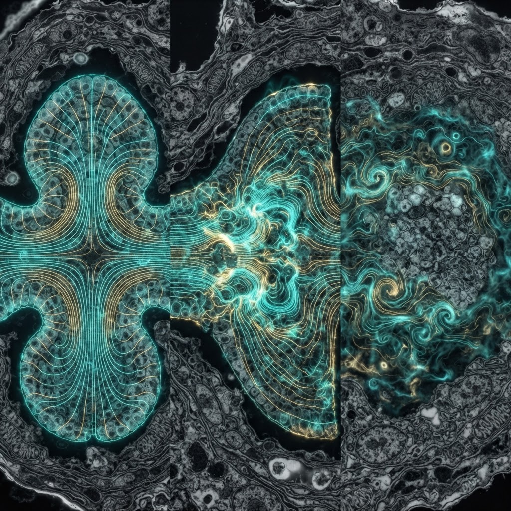
Two canonical patterns emerge. First, the dominant dipolar pattern tied to ventricular depolarization is highly coherent across sensors at the heartbeat fundamental frequency and its harmonics, producing reproducible spatial maps of the cardiac vector. Second, finer-grained coherence can reveal transient regional desynchronization or re-entrant activity in arrhythmias, where local fields lose phase alignment with the global cardiac cycle. High-resolution MCG can therefore distinguish globally coherent contractions from pathological pockets of incoherence.
Physiological entrainment: mechanisms and limits
The term entrainment covers several related phenomena: one oscillator adjusting its phase or frequency to another, and two systems becoming phase-locked. In the cardio-respiratory domain, entrainment is commonly mediated by neural and mechanical pathways (baroreflexes, vagal modulation, thoracic pressure changes). The question addressed here is whether the heart's electromagnetic field itself can contribute to entrainment of nearby excitable tissue or systemic rhythms.
From a biophysical standpoint, direct electromagnetic entrainment by the heart is constrained by field amplitude and coupling. The picoTesla-level fields generated externally are several orders of magnitude weaker than thresholds typically required to drive membrane polarization in isolated excitable cells when applied as uniform fields. However, there are plausible scenarios where field-mediated effects could matter: near-field exposure (very close proximity, such as implanted sensors or adjacent tissue), resonant interactions at specific frequencies, or collective behavior in populations of cells that amplify weak synchronizing inputs.
Importantly, many reported correlations between cardiac phase and other physiological rhythms are more parsimoniously explained by autonomic, hemodynamic, or mechanotransductive coupling than by direct electromagnetic forcing. That said, controlled in vitro and computational studies show that under specific parameter regimes weak oscillatory magnetic fields can bias ion channel gating and spike timing, suggesting that electromagnetic entrainment is not impossible — only typically small compared with other coupling modalities in vivo.
Implications and next steps
MCG's unique sensitivity to current orientation and its improving spatial fidelity make it a valuable tool for studying cardiac coherence and its breakdown in disease. For clinical translation, mapping coherence patterns could refine arrhythmia localization, guide ablation therapy, and track recovery of coordinated contraction after infarction.
For fundamental physiology, the key questions remaining are quantitative. How large are field-mediated effects relative to autonomic and mechanical coupling in intact organisms? Under what spatial and spectral conditions can cardiac magnetic fields modulate nearby excitable tissues? Answering these requires combined approaches: high-sensitivity MCG, close-proximity sensor arrays (including wearable OPMs), targeted in vitro experiments, and biophysically detailed modeling to quantify plausible field–tissue coupling.
Bottom line: Magnetocardiography documents robust, spatially organized cardiac magnetic coherence at picoTesla amplitudes. Direct electromagnetic entrainment of other physiological rhythms is physically constrained and likely a minor player compared with neural and mechanical pathways, but it remains a scientifically tractable mechanism under specific near-field or resonant conditions worthy of rigorous follow-up.