Bioelectricity resurfaces as a central morphogenetic force
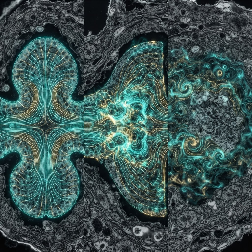
For decades morphogenesis has been narrated in chemical terms: gradients of diffusible morphogens, transcriptional networks and cell mechanics. A quieter story — cells' native electrical states — has been gaining clarity and traction. Endogenous bioelectric fields, arising from transmembrane potentials, ion channels and gap-junctional currents, are now recognized as instructive regulators of cell behavior across development, regeneration and disease.
What the evidence shows
Work spanning planaria, amphibian embryos and mammalian tissues demonstrates that spatial patterns of membrane voltage (Vmem) carry information that predicts and instructs anatomical outcomes. Experimental manipulations that alter regional Vmem — by pharmacology, ion-channel misexpression or light-activated channels — can change cell proliferation, migration, and differentiation programs and redirect where structures form. In regenerating systems, transient electric cues can re-specify polarity and scale of regrown tissues; in embryos, defined voltage domains precede gene-expression changes responsible for organogenesis.
Mechanisms: how voltage talks to genes
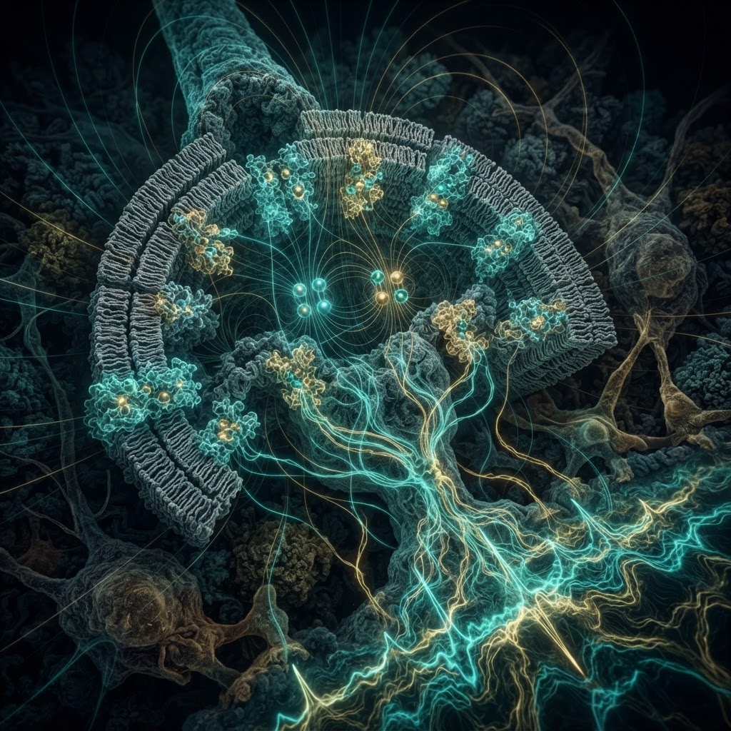
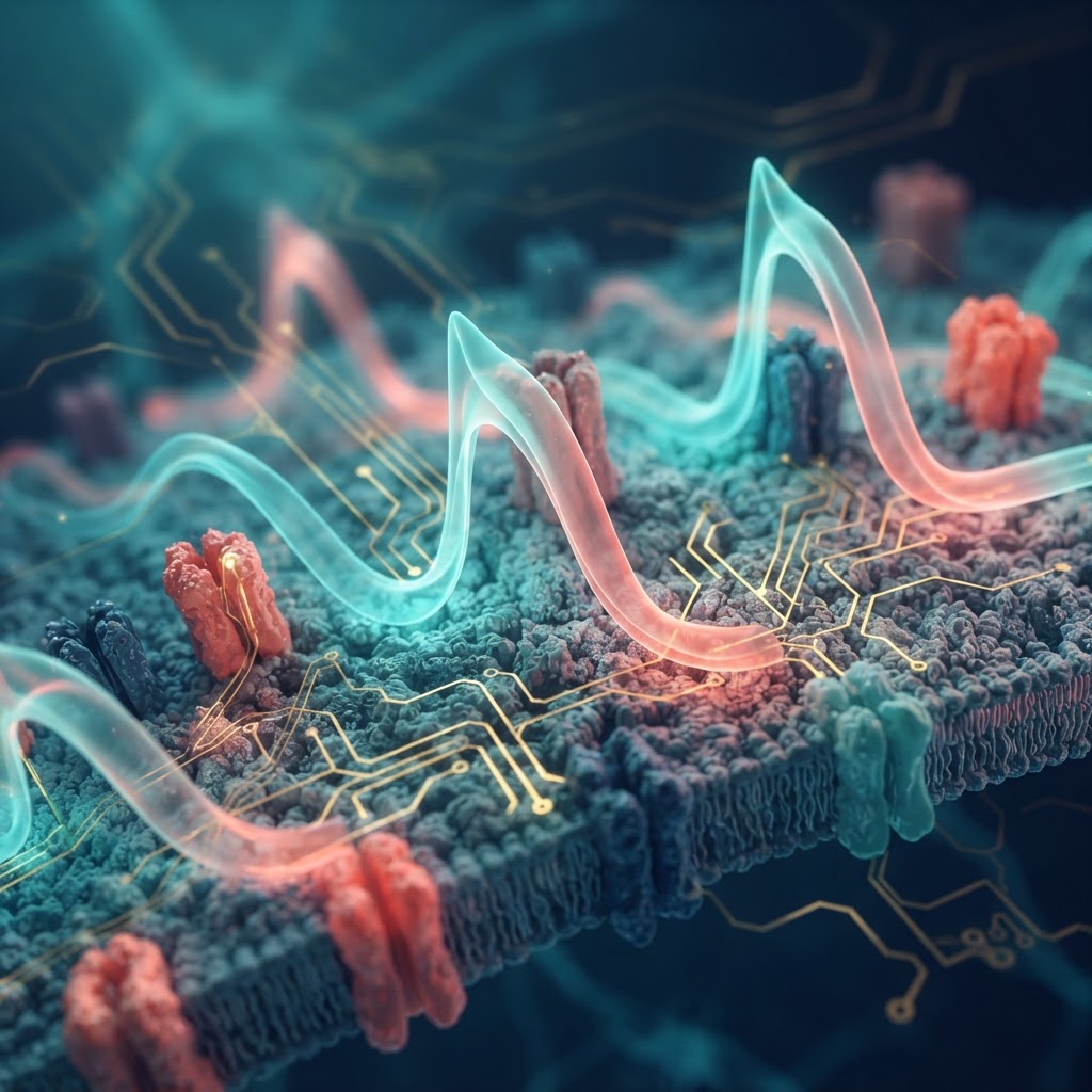
Voltage is not a mystical language: it funnels through biochemical mediators. Changes in Vmem regulate the flow of calcium, protons and other second messengers, alter transporter activity, and influence the activity of voltage-sensitive signaling proteins. These intermediaries then impinge on canonical transcriptional and epigenetic machinery. Gap junction networks convert single-cell electrical states into tissue-scale patterns, producing robust, reproducible voltage maps that coexist with morphogen gradients.
Tools that made the case
- Optogenetics and chemogenetics: enable temporally precise depolarization/hyperpolarization in specific cell populations.
- Voltage-sensitive dyes and genetically encoded voltage indicators: provide spatial maps of Vmem during development and repair.
- Ion-channel engineering: allows synthetic reprogramming of tissue electrical states to test sufficiency for patterning outcomes.
Where bioelectricity matters now
Beyond academic interest, bioelectric control is a practical lever. In regenerative medicine researchers are exploring electrical interventions to coax tissues toward desired forms; in oncology, disrupted bioelectric circuits correlate with tumorigenic transformation and could become diagnostic or therapeutic targets. Because electrical cues integrate over many cells and span large distances rapidly, they provide a complementary axis of control to molecular signals that diffuse or act locally.
Outstanding debates and technical hurdles
Key uncertainties remain. How to read the bioelectric code — which patterns are meaningful and which are epiphenomena — is unresolved. Quantitative models that link measured Vmem maps to downstream gene regulatory changes are still emerging. Measurement challenges persist: many voltage-sensitive tools trade spatial resolution for sensitivity, and in vivo mapping at cellular resolution across whole tissues is technically taxing. Finally, the mechanistic chain from ion flux to durable morphological change needs further dissection to pinpoint therapeutic entry points.
Looking ahead
The field is converging on a synthesis: bioelectric states are a parallel information channel in morphogenesis, acting together with biochemical gradients and mechanical cues. Decoding that channel offers a new set of design rules for engineering tissue form and for intervening in regeneration and cancer. The near-term goal is pragmatic — generate predictive, testable models that let researchers set electrical boundary conditions and reliably produce desired anatomical outcomes.
Bioelectric fields are not a mysterious alternative to molecular biology — they are an integrated control layer whose practical utility is just beginning to be realized.