Under the skin, a quiet current runs
When you cut yourself the story most people are told is chemical: clotting factors, growth factors, immune cells and a cascade of gene expression. That story is true — but incomplete. Less visible and rarely cited in mainstream accounts is a physical language layered on top of biochemistry: endogenous voltage gradients. Across epithelia and damaged tissues, small but organized voltage differences — millivolts per millimeter — act as a directional scaffold for cells and a patterning signal for tissues.
How wounds make electricity
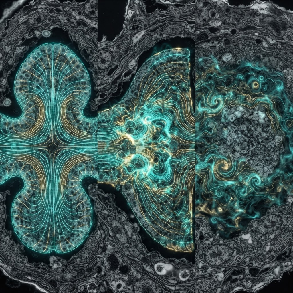
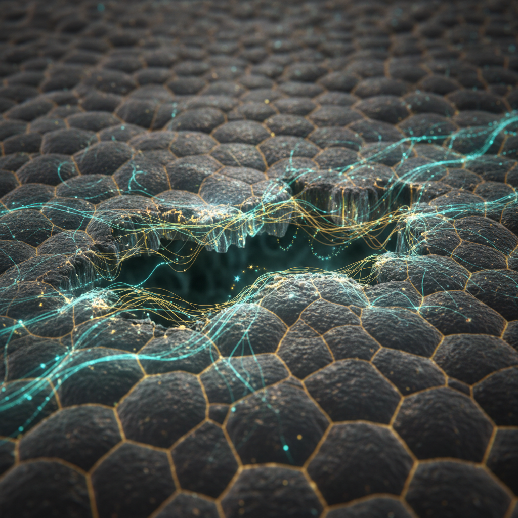
Epithelia maintain a transepithelial potential (TEP) by polarized ion transport: pumps, channels and tight junctions keep the inside of a tissue at a different potential than its surface. A breach collapses that local TEP, and current flows from intact regions into the wound. The result is an outward-directed electric field centered on the lesion. Measured fields are tiny by engineers' standards (tens to hundreds of mV/mm), but they are neither trivial nor noisy: they are sustained, directional and spatially patterned.
Cells respond — predictably and usefully
Many cell types — keratinocytes, fibroblasts, endothelial cells, neurons and immune cells — detect these fields and migrate directionally, a behavior called galvanotaxis or electrotaxis. In amphibians and planarians, altering endogenous voltages changes regeneration outcomes: eyes, tails or mis-patterned heads can be induced or suppressed by tuning membrane potentials or ion flows. Blocking ion channels, pharmacologically or genetically, distorts wound currents and impairs repair. Conversely, externally applied microcurrents or electrodes can accelerate closure and bias cell orientation.
Why this is overlooked
Two cultural biases are at work. Molecular biology prizes soluble ligands and genetic circuits; physical cues like voltage and current are harder to fit into gene-first narratives. Second, the measurements are technically finicky — you need vibrating probes, microelectrodes, voltage-sensitive dyes and careful geometry to map fields reliably. When good measurements are scarce, fields sit in the blind spot between electrophysiology and developmental biology.
Mechanisms — known and mysterious
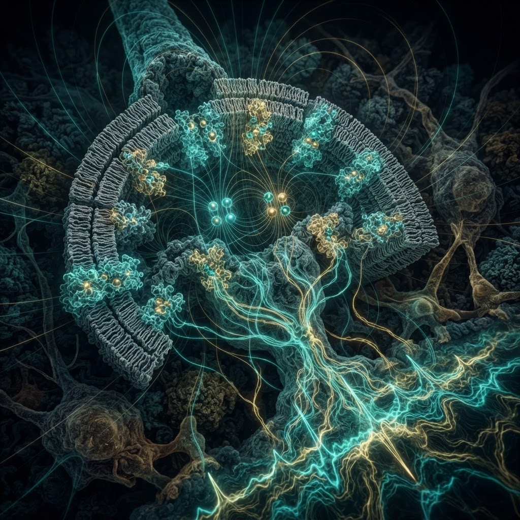
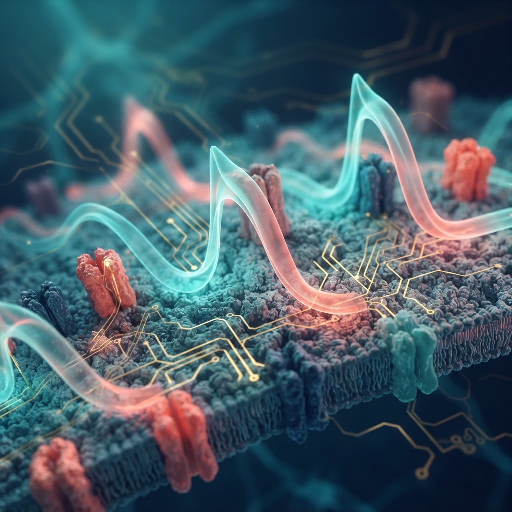
Known: ion pumps (Na+/K+-ATPase), channels (ENaC, HCN, various voltage-gated channels), and gap junctions establish and propagate bioelectric states. Calcium signaling and second-messenger pathways like PI3K/PTEN mediate directional responses in many cell types, linking membrane potential (Vmem) change to cytoskeletal remodeling.
Mysterious: how do graded Vmem patterns encode complex anatomical instructions? Hypotheses include electrophoretic redistribution of charged morphogens, voltage-dependent conformational changes in membrane proteins, and Vmem control of gene transcription via voltage-sensitive transcriptional regulators. The spatiotemporal code — how a Vmem map turns into a reproducible organ or coordinate axis — remains poorly defined.
Why it matters
- Practical: modest electric stimulation is already used in wound-care devices; a deeper biophysical understanding could rationalize dosing, timing and electrode design.
- Theoretical: bioelectric patterns act upstream of gene expression in some contexts — a non-genetic information layer for anatomy.
- Translational: ion channel modulators and targeted bioelectric interventions could become regenerative medicine tools.
Where to look next
We need better spatiotemporal maps of Vmem during healing, molecular readouts of voltage translation, and integrative models that couple electrophysiology to gene-regulatory networks. The gap is not that the fields are weak, but that our conceptual toolkit has undervalued directional, physical cues. Reconciling bioelectricity with mainstream molecular narratives could unlock low-energy, precision therapies for chronic wounds and organ regeneration.
Wounds are not merely biochemical crises; they are electrical events with instructions. If we learn to read and write those instructions, the clinical payoff could be quietly enormous.