Summary
Over the past decade, a steady accumulation of experimental and theoretical work has shifted bioelectricity from a niche curiosity to a mainstream candidate mechanism for morphogenesis. Endogenous transmembrane voltage gradients (Vmem), patterns of ion transporter activity, and intercellular electrical coupling now appear to carry positional information and to bias gene-regulatory networks during development, regeneration, and disease. New tools for measuring and manipulating membrane voltage in vivo have been decisive: genetically encoded voltage indicators, optogenetic ion pumps, and precise ion-channel perturbations let researchers test causality rather than rely on correlation.
What’s changed recently
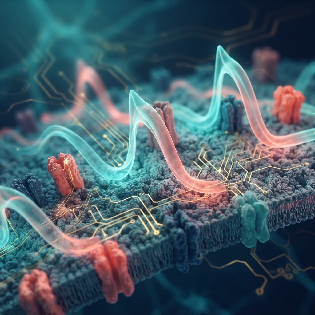
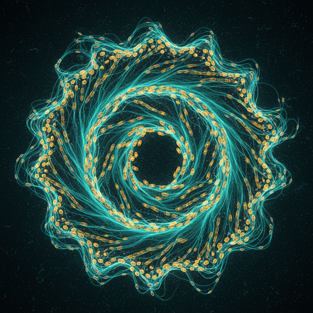
Measurement and perturbation are converging. Genetically encoded voltage indicators (GEVIs) such as ArcLight derivatives and faster probes paired with high-speed imaging now reveal voltage domains at cellular and tissue scales during embryogenesis. Optogenetic actuators and targeted delivery of ion-channel constructs allow researchers to reconfigure those domains with spatiotemporal precision. Several groups have used these approaches to show that altering Vmem in localized regions can redirect tissue identity, induce ectopic organogenesis, or rescue regenerative failures in model systems including Xenopus, planaria and mammalian cell systems.
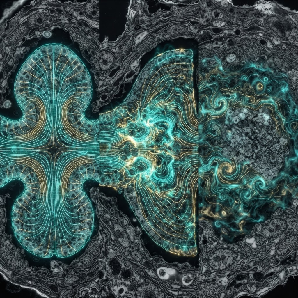
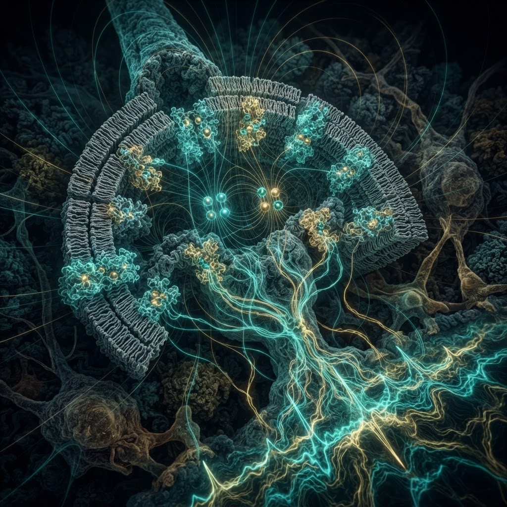
Mechanistic links are sharpening. Voltage changes propagate through gap-junctional networks and can gate downstream biochemical pathways: voltage-sensitive calcium entry, pH shifts, and the activity of voltage-sensitive phosphatases and transporters provide plausible transduction routes to transcriptional circuits. Computational models that couple electrophysiology, diffusion and gene regulation are beginning to reproduce experimentally observed pattern outcomes, supporting the claim that bioelectric states store and transmit patterning information beyond local genetic expression.
Why this matters now
Bioelectric cues offer a layer of control that is fast, reversible, and spatially reconfigurable — properties that genetic or chemical signals lack. For regenerative medicine, that means the potential to instruct tissues to rebuild correct size and shape without introducing new cell types or permanently altering genomes. In developmental biology, recognizing voltage as an instructive signal forces a re-evaluation of how positional information is encoded and integrated.
Active debates and open challenges
- Causation vs. correlation at scale. While local manipulations have compelling effects, it remains difficult to prove that naturally occurring bioelectric patterns are necessary and sufficient for large-scale organ patterning in higher vertebrates.
- Transduction specificity. Multiple biochemical routes link Vmem to downstream effectors — identifying which are decisive in each context is ongoing work.
- Standardization and reproducibility. Measurements of absolute Vmem in tissues are technically challenging and lab-to-lab variance persists. The field needs shared benchmarks for probes, perturbations and analytical pipelines.
Outlook
Expect rapid progress: improved voltage reporters, synthetic bioelectric circuits, and refined computational frameworks will coalesce into testable rules for electrical patterning. Translation to therapeutics — wound healing, organoid engineering, and targeted anti-cancer strategies that restore proper morphogenetic fields — is plausible within a decade, but will require rigorous mechanistic validation and careful assessment of off-target physiological effects.
Bioelectricity is no longer a marginal curiosity; it is an interfacial language between ion physics and gene regulation, and decoding it may rewrite how we think about form.