The Physical Reality of Ultraweak Photon Emission
At a baseline metabolic state, living mammalian tissue emits a steady flux on the order of 10 to 100 photons per second per square centimetre (measured; interpretation contested). This flux spans a broad spectral range from roughly 200 to 800 nanometres, encompassing both ultraviolet and visible wavelengths. The phenomenon, ultraweak photon emission (UPE), is not an artifact of ambient light leakage; it is recorded by photomultiplier tubes and cooled-CCD arrays inside heavily shielded, light-tight chambers that exclude every other photon source in the room.
The detection problem is the real bottleneck, and it is a counting problem before it is a biological one. At room temperature a photocathode generates its own dark counts from thermionic emission, and when that dark-count rate sits within an order of magnitude of the biological signal, the measured value becomes the small difference between two noisy Poisson processes, each carrying its own square-root uncertainty. Cooling the detector to tens of degrees below zero suppresses thermionic noise by a substantial factor, but cosmic-ray muons still strike the photocathode at intervals and deposit pulses of anomalously high amplitude that must be identified and vetoed by pulse-height discrimination or coincidence logic, or they inflate the apparent biological flux. The remaining strategy is brute integration time: because the fractional uncertainty in a Poisson count falls only with the square root of total counts accumulated, resolving a 10-photon-per-second signal to useful precision can require minutes to hours of continuous dark-adapted acquisition, during which the sample's own metabolic state must not drift (Liebert 2025, Sci Rep).
This is also why reported flux values differ across laboratories by an order of magnitude or more, and why that spread should not be read as evidence against the phenomenon. Detector quantum efficiency, dark-count rate, cooling temperature, collection solid angle, spectral bandpass, contact versus non-contact geometry, and the metabolic state and species of the tissue sample all shift the measured number independently of any underlying biological reality. An experiment that integrates over a wide visible-plus-UV window at close geometric coupling will report a higher flux than one that filters to a narrow visible band at a distance, even from identical tissue.
The dominant and well-supported mechanism for this emission is oxidative: the recombination of reactive oxygen species (ROS) and the relaxation of electronically excited carbonyl and singlet-oxygen species generated during lipid peroxidation in mitochondria and cellular membranes (established). Because these excited states relax through specific, chemically determined transitions rather than a smooth thermal continuum, the emission spectrum should in principle resolve into discrete bands layered on a broad background, a signature that distinguishes biochemical luminescence from blackbody-like noise. The intensity scales directly with oxidative stress, and that relationship is quantitative enough that increased stress produces predictable patterns of decay in the emitted intensity, allowing researchers to use UPE as a label-free metabolic readout (Hideg 2022, Sci Rep).
That oxidative-origin logic generates testable predictions rather than settled facts about specific physiological states. During sustained exercise, mitochondrial oxygen flux rises, so UPE should rise concurrently and track recovery kinetics after exertion ends. During ischaemia, oxygen deprivation should initially suppress the signal, but the reperfusion that follows is expected to produce a transient burst as the oxidative machinery re-engages abruptly against a backlog of reduced substrates. In cell death, mitochondrial permeability transition and the associated ROS burst predict a spike in emission that precedes or accompanies the terminal stages of apoptosis, followed by a collapse as the cell's oxidative chemistry disintegrates. None of these are new mechanisms; they are direct, falsifiable consequences of treating UPE as an oxidative byproduct rather than an exotic information channel.
The Coherence Hypothesis and the Measurement Deficit
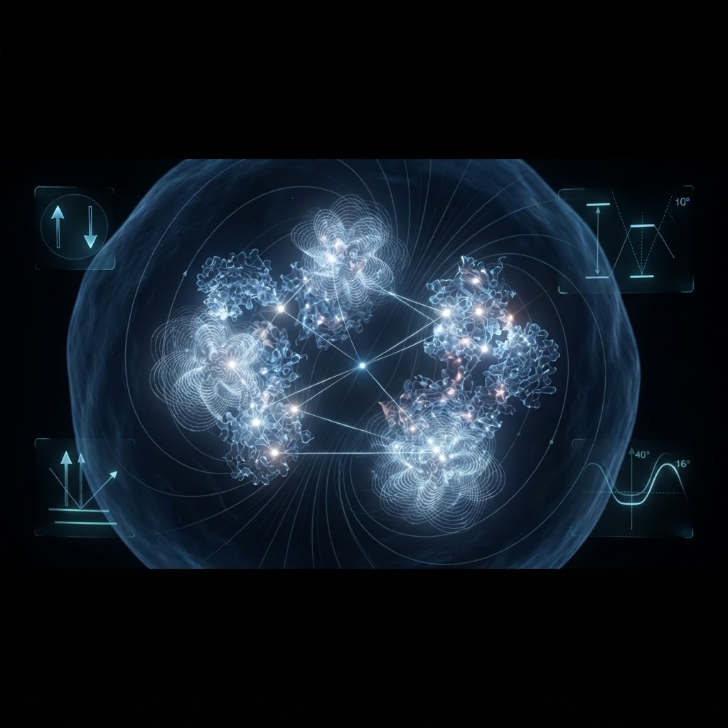
The literature surrounding these photons is fractured by a persistent claim: that this light is not merely thermal noise, but constitutes a coherent biophoton field capable of orchestrating cellular processes. This is fundamentally a claim about photon statistics, yet almost no experiment in the biological literature is actually designed to test it. Distinguishing a coherent or squeezed field from a chaotic thermal source at these extreme low intensities requires measuring the second-order correlation function, g²(0), and most biological detectors integrate over macroscopic ensembles of emitters, a process that mathematically washes out the very photon-by-photon statistical signature that would prove coherence (model-dependent).
Squeezing and coherence have been claimed in the literature, but the claims rest on indirect proxies rather than a direct correlation measurement. Some researchers infer coherence from the conditional probability of zero-photon detection events, a quantity that can be suggestive but does not by itself establish sub-Poissonian statistics with the rigor the physics community expects (Popp 2022, Springer). Consequently, the broader consensus remains that no reliable evidence for coherence or non-classicality of UPE has been achieved through current methodologies (Brizhik 2015, arXiv).
The Coherence Test, Done Properly
A decisive g²(0) measurement requires a Hanbury Brown and Twiss arrangement: the biological photon stream is split by a beamsplitter onto two independent single-photon detectors, and the rate of genuine coincidences is compared against the accidental coincidence rate predicted by Poisson statistics for the measured mean intensity. Antibunching, g²(0) below 1, shows up as a coincidence deficit relative to that chance-level prediction, and detecting it with statistical confidence requires enough total counts that the shot-noise error on the coincidence estimate shrinks below the size of the deficit itself.
At a collected rate of a few photons per second after accounting for limited numerical aperture and finite detector efficiency, reaching that confidence threshold while also subtracting dark counts and cosmic-ray events cleanly demands integration windows of many hours, far longer than most living tissue preparations remain in a stable, defined metabolic state. The instrument also needs ultra-low dark-count single-photon avalanche diodes or cooled photomultipliers whose intrinsic noise floor sits well below the biological signal, plus tight, high-efficiency collection optics to raise the detected rate enough that the measurement finishes before the sample itself changes. This is a hard experiment, but it is hard in a specific, buildable way, not an unsolvable one.
One of the most robust and reproducible phenomena in this domain is delayed luminescence. When biological tissue is illuminated and then placed in darkness, the subsequent photon emission decays not exponentially, but hyperbolically, and that shape is stable across widely different tissues and organisms. This is physically significant because a hyperbolic decay is mathematically a superposition of many exponential decays spread across a broad, continuous distribution of relaxation times, rather than the signature of a single dominant fluorophore relaxing at one fixed rate. That same logic, a broad spread of relaxation timescales superposing into a single aggregate signal, is the mechanism invoked to explain 1/f noise spectra elsewhere in biological dynamics, which suggests cellular architecture behaves as a highly coupled, dispersive optical medium rather than a simple point emitter.
Neural Signaling and the Waveguide Constraint
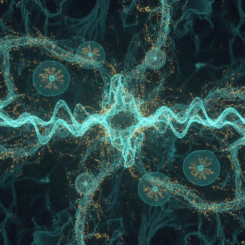
The neural-signaling claim, that biophotons serve as a conduction mechanism or information channel in nerves, represents the most aggressive interpretation of UPE (speculative — flagged). Proponents argue that the myelin sheath could act as an optical waveguide, transmitting photons between nodes of Ranvier. While correlations between UPE and electroencephalographic activity have been reported, these studies frequently suffer from tiny sample sizes, inadequate dark-count subtraction, and detection geometries that cannot distinguish brain emission from scalp emission, and recent analysis suggests that extracranial UPE signals are likely artifacts of background light and scalp tissue rather than neural activity itself (Salari 2026, arXiv).
A decisive experiment would need to satisfy four conditions simultaneously: spatial resolution fine enough to isolate single neurons or axon segments rather than tissue volumes; single-unit electrophysiological recording run concurrently with photon counting so a spike can be time-locked to a photon event; replication across independent laboratories with blinded analysis; and a fully characterized optical path with independently measured scattering and absorption coefficients ruling out stray light. Current baseline measurements, including population-level neural-cell emission rates around 11 counts per second, satisfy the metabolic-baseline requirement but not the single-unit spatial resolution requirement, since they average over many cells and cannot be time-locked to individual action potentials (Liebert 2025, Sci Rep). The deeper obstacle is physical rather than instrumental: thermal fluctuations and high-frequency biological noise severely limit how long any hypothesized quantum state could propagate coherently across even a single cell's dimensions, let alone between neurons (Pospisil 2026, Prog Biophys Mol Biol).
The Arithmetic of Neural Biophoton Signalling
Start with the measured baseline: roughly 10 photons per second per square centimetre at the low end of reported flux. Converting that surface rate to a volumetric emission for a cubic millimetre of tissue, treating the volume as uniformly and isotropically emitting, yields on the order of 10⁴ photons per second from that small block of tissue. Apply a typical biological fluorophore quantum yield of about 10⁻³, which already discounts most emitted energy to non-radiative relaxation before a photon escapes at all.
Now apply attenuation: lipid-rich brain tissue scatters and absorbs visible light heavily, so even a path length of a few hundred micrometres removes the large majority of the remaining photons before they reach a neighbouring cell. Carry that surviving fraction forward to the timescale that matters for signalling, the roughly one-millisecond window of an action potential, and the expected number of photons arriving at a specific neighbouring target in that window drops far below one. For reliable detection, the system either needs to fire photons in gross redundancy, many orders of magnitude more than the baseline flux, or needs the receiving structure to sit at a synaptic-cleft-scale distance of tens of nanometres rather than cellular-scale micrometres. Named plainly: the assumptions are baseline flux, quantum yield, attenuation over distance, and the millisecond integration window of a spike, and each one independently pushes against long-range optical signalling.
| Parameter | Chemiluminescence Control | Observed Biophoton Emission | Theoretical Coherent Source |
|---|---|---|---|
| Flux (photons/s/cm²) | 10^4 - 10^6 | 10 - 100 | Variable, but requires threshold |
| Decay Profile | Exponential | Hyperbolic | Phase-locked / Squeezed |
| Photon Statistics | Poissonian (Thermal) | Largely Poissonian (Ensemble) | Sub-Poissonian (g²(0) < 1) |
| Origin | Specific chemical reaction | Lipid peroxidation / ROS | Coupled oscillator network |
The verdict on ultraweak photon emission is clear: it is a real, measurable signal rooted in oxidative metabolism, with a spectral and kinetic structure that follows directly from that chemistry. It is an unproven communication channel, and the claims of quantum coherence remain unsupported by the necessary statistical measurements, not because the question is unanswerable but because the decisive instrument has not been pointed at it long enough. The Hanbury Brown and Twiss adaptation described above is difficult, expensive in integration time, and unglamorous, but it is not beyond current optical engineering; it is a cheap experiment, in instrumentation terms, that remains unbuilt, leaving the field to cycle through theoretical models without the anchor of hard quantum-optical data. This absence of rigorous statistical measurement bridges directly into the broader investigation of biological noise and 1/f dynamics, where the structure of fluctuation, not the mean signal, so often carries the information.
What would change our mind
- A replicated measurement of the second-order correlation function g²(0) from a single biological emitter, integrated long enough to statistically exclude Poissonian thermal statistics, showing sub-Poissonian photon counting at a confidence level accepted in quantum optics generally.
- In vivo demonstration of optical waveguiding in mammalian axons, with independently measured attenuation coefficients low enough to permit single-photon transmission over millimetre distances rather than tens of nanometres.
- A spatially resolved, single-unit recording showing deterministic, phase-locked photon emission time-locked to an individual action potential, replicated across independent laboratories, and shown to be independent of generalized oxidative stress rather than a byproduct of it.