A Skeleton That Speaks in Voltage
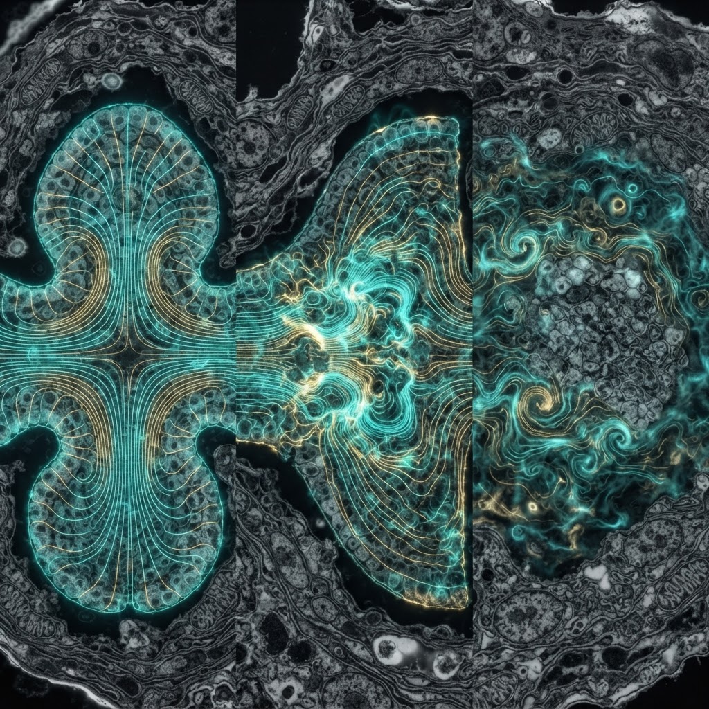
When a runner's foot strikes pavement, the impact doesn't merely compress bone — it polarizes it. Collagen fibers and hydroxyapatite crystals within the bone matrix deform under load, and that deformation displaces bound charges, generating transient electrical potentials measurable in the range of 1–100 millivolts across cortical bone surfaces. This is the piezoelectric effect — the conversion of mechanical stress into electrical polarization — and in bone it is not an epiphenomenon. It is a primary language through which the skeleton encodes mechanical experience and directs cellular response.
The concept has been refined over six decades of biophysics, from mid-20th-century curiosities about dry bone specimens to contemporary work resolving the nanoscale electromechanical behavior of individual collagen fibrils. Yet despite this history, the field remains vigorously contested. The precise molecular transducers, the relative contributions of piezoelectricity versus streaming potentials, and the downstream signaling cascades linking voltage to gene expression remain subjects of active and sometimes acrimonious debate.
Historical Foundations: Fukada, Yasuda, and the Bone Voltage Discovery
The modern story begins in 1957, when Japanese biophysicists Eiichi Fukada and Iwao Yasuda published their landmark measurements of piezoelectric potentials in dry cortical bone. Using a Rochelle salt piezoelectric standard for comparison, they demonstrated that compressed bone produced voltage outputs consistent with a piezoelectric coefficient on the order of 0.7 pC/N — modest by crystal standards, but physically meaningful in a biological context. Yasuda had already suspected a connection: his earlier clinical observations suggested that mechanical stress governed the directionality of bone growth, an intuition later formalized as Wolff's Law in biomechanical terms.
The critical conceptual leap was recognizing that bone's piezoelectricity resided primarily in its collagen component rather than in the inorganic hydroxyapatite phase. Dry collagen is a non-centrosymmetric polymer — its triple-helix structure lacks an inversion center — which is the prerequisite for piezoelectricity at the molecular level. Subsequent work in the 1960s and 70s confirmed that demineralized bone retained piezoelectric activity, while denatured collagen lost it, firmly localizing the effect to the organic matrix.
Molecular Mechanisms: Collagen, Hydroxyapatite, and the Composite Architecture
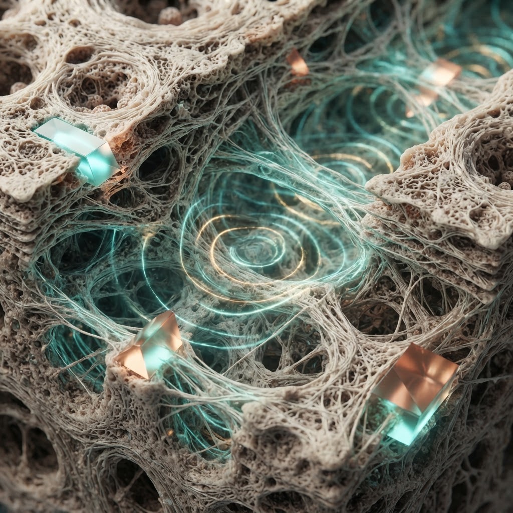
Contemporary understanding treats bone's electromechanical behavior as emerging from a hierarchical composite at multiple length scales. At the nanoscale, type I collagen fibrils — the dominant organic component of bone — exhibit piezoelectric coefficients that have been directly measured by piezoresponse force microscopy (PFM) in the range of 1–10 pm/V. The asymmetric arrangement of peptide dipoles along the collagen triple helix, combined with the ordered alignment of fibrils in lamellar bone, creates a macroscopic piezoelectric response that is both magnitude- and direction-sensitive.
The hydroxyapatite (HAp) crystals embedded within and between collagen fibrils add complexity. Stoichiometric HAp has a hexagonal crystal structure with limited piezoelectricity, but biological HAp is non-stoichiometric, carbonate-substituted, and nanometer-scaled — conditions that introduce lattice asymmetries potentially amplifying the electromechanical response. Recent molecular dynamics simulations suggest that the collagen-HAp interface itself may be a zone of enhanced electromechanical coupling, where protein-crystal interactions constrain fibril deformation in ways that maximize charge separation.
A critical distinction must be drawn between true piezoelectricity and streaming potentials, which have been conflated in much of the older literature. Streaming potentials arise when fluid — interstitial fluid within the lacunar-canalicular network — is driven through charged pore surfaces by mechanical pressure gradients. Both mechanisms generate electrical signals in loaded bone, but they differ in temporal signature (streaming potentials decay with fluid redistribution on timescales of seconds to minutes), spatial distribution, and cellular accessibility. Most current models treat them as complementary rather than competing mechanisms, with piezoelectric signals dominating at high-frequency loading and streaming potentials more relevant to sustained compressive loads.
Cellular Transduction: How Osteocytes Decode the Electric Signal
The mechanosensory cell of bone is the osteocyte — a terminally differentiated osteoblast entombed within the mineralized matrix, connected to neighbors and to surface cells via a vast dendritic network threading through canaliculi. With approximately 42,000 osteocytes per cubic millimeter of cortical bone in humans, this network constitutes a sensory syncytium of extraordinary density. The question of how electrical signals reach and activate these cells is not fully resolved, but several mechanisms have strong experimental support.
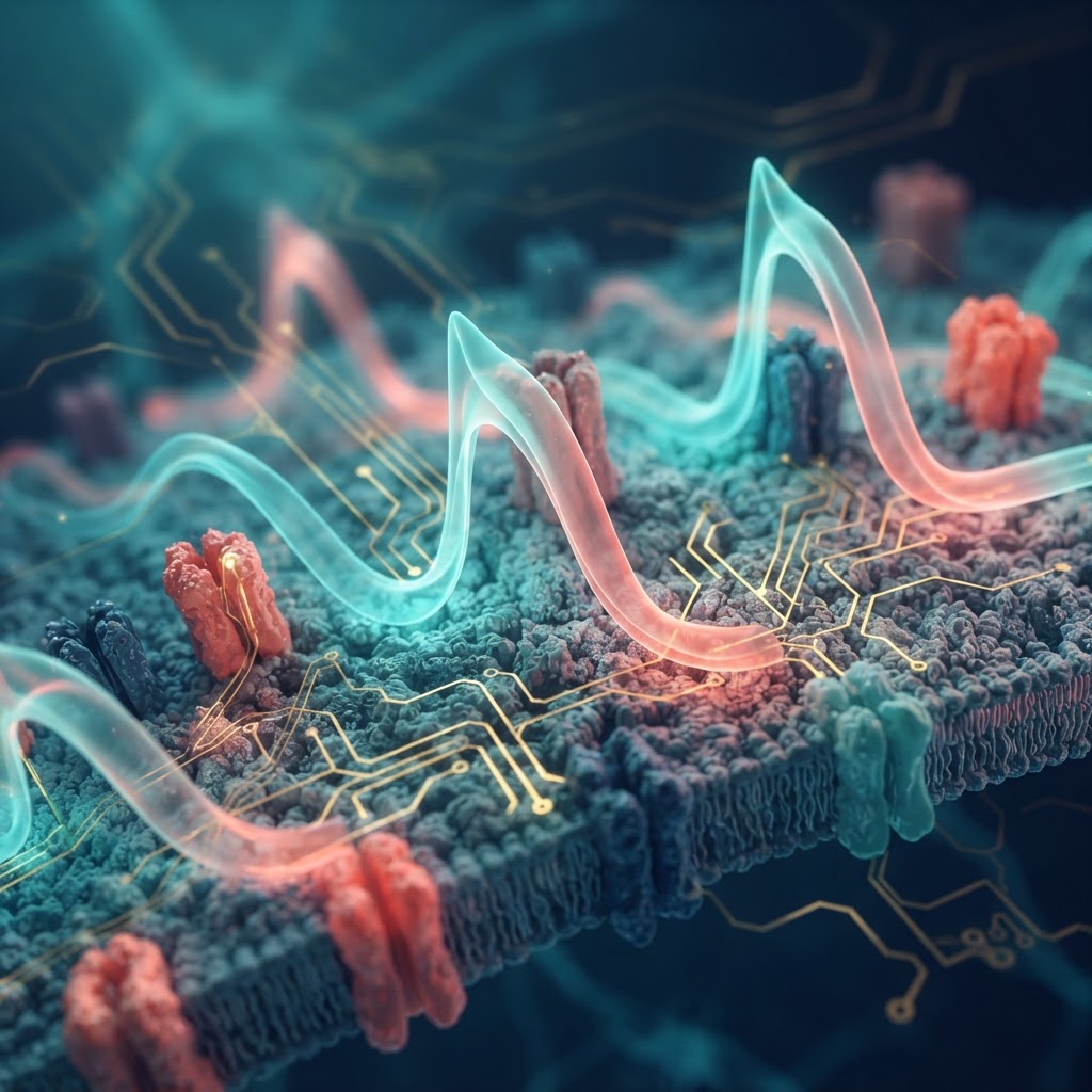
Voltage-gated ion channels, particularly L-type calcium channels (Cav1.2), are expressed on osteocyte membranes and respond to membrane depolarization with calcium influx. Piezoelectrically generated fields — even if attenuated by tissue conductivity — can create local voltage gradients sufficient to open these channels, triggering intracellular calcium transients that propagate through the network via gap junctions (primarily Connexin 43). This calcium wave is itself a secondary messenger cascade, activating nitric oxide synthase, prostaglandin E2 release, and ultimately modulating the RANK/RANKL/OPG axis that controls osteoclast-osteoblast balance.
Separately, evidence from cell culture studies using applied electric fields in the physiologically relevant range (10–1000 mV/mm) demonstrates direct effects on osteoblast proliferation, differentiation, and matrix synthesis. Applied fields of this magnitude upregulate BMP-2, osteocalcin, and alkaline phosphatase expression, consistent with a pro-anabolic role for the electrical microenvironment. Critically, these effects are frequency-dependent: sinusoidal fields at 20 Hz — approximating walking frequency — produce stronger osteogenic responses than static fields, suggesting that the cellular machinery is tuned to dynamic loading signatures.
Competing Hypotheses and Methodological Fault Lines
The field is haunted by a persistent methodological challenge: measuring endogenous piezoelectric fields in living, hydrated bone is extraordinarily difficult. Most classical measurements were performed on dry bone specimens, which may overestimate in vivo piezoelectric contributions because hydration reduces the piezoelectric coefficient of collagen by partially screening molecular dipoles. More recent in situ measurements using implanted microelectrodes and optical voltage indicators suggest that stress-generated potentials in living bone are smaller than early estimates — potentially in the range of 0.1–10 mV at the cellular scale — raising questions about whether they are sufficient to directly gate ion channels or whether amplification mechanisms are required.
One prominent competing hypothesis holds that mechanical deformation of the osteocyte cell body and dendrites — not electrical signaling — is the primary transduction mechanism, mediated by integrin-cytoskeleton coupling, primary cilia deflection, and stretch-activated channels such as Piezo1 and TRPV4. Knockout studies removing Piezo1 from osteoblast lineage cells produce dramatic deficits in load-induced bone formation, arguing that this mechanically gated ion channel is indispensable in a way that pure piezoelectric signaling cannot substitute for. The resolution may lie in recognizing that electrical and purely mechanical transduction are not mutually exclusive: they may operate at different timescales and spatial scales, with the electrical signal providing a long-range, rapid-onset cue that is locally amplified by direct mechanical stimulation of cells in the most highly strained regions.
Clinical Implications and Therapeutic Horizons
The biomedical stakes of understanding bone piezoelectricity are considerable. Osteoporosis — affecting an estimated 200 million people globally — is fundamentally a failure of mechanosensory feedback: the skeleton loses its ability to match bone formation to mechanical demand. If piezoelectric or electrically-mediated signals are disrupted by aging-related changes in collagen crosslinking, crystal stoichiometry, or osteocyte connectivity, therapeutic restoration of the electrical microenvironment becomes a rational target.
Externally applied electrical stimulation via implanted electrodes or transcutaneous devices has been used clinically since the 1970s, primarily for fracture non-unions. The FDA-approved devices deliver direct current or pulsed electromagnetic fields, and while their clinical efficacy is established for certain indications, the mechanistic basis remains poorly characterized. More recent approaches include piezoelectric scaffolds for bone tissue engineering — materials such as polyvinylidene fluoride (PVDF), barium titanate nanoparticles, and piezoelectric biopolymers that generate endogenous charge under cyclic loading in vitro and in vivo, promoting osteogenic differentiation without exogenous electrical sources. Early animal studies show promise, but translation to human use remains in early phases.
Emerging nanotechnology approaches propose embedding piezoelectric nanoparticles directly into defect sites, where physiological loading would autonomously drive local electrical stimulation — a kind of self-powered bone regeneration that exploits the same physics evolution spent hundreds of millions of years optimizing.
Open Questions and the Road Ahead
For all its history, bone piezoelectricity research sits at a productive frontier. Key unresolved questions include: What is the actual in vivo electric field magnitude experienced by osteocytes during normal locomotion? High-resolution voltage imaging in living bone, perhaps using genetically encoded voltage indicators expressed in osteocytes, may finally answer this. How does the piezoelectric response change with aging, disease, and disuse atrophy — and do these changes causally contribute to skeletal fragility? What are the precise gene regulatory targets of electrically stimulated osteocytes, and how do they integrate with biochemical signals like IGF-1, sclerostin, and parathyroid hormone?
Perhaps most fundamentally: is bone's piezoelectricity a designed feature or an evolutionary spandrel — a structural consequence of collagen's asymmetric chemistry that biology subsequently co-opted for signaling? The answer may be both. What is certain is that the skeleton is not a passive structural scaffold. It is an electroactive tissue, perpetually parsing the language of force into the vocabulary of charge, and using that information to rebuild itself accordingly.