Executive hypothesis
Biological structures at physiological temperature can host measurable quantum-coherent spin or excitonic dynamics with correlation times from tens of nanoseconds to microseconds — detectible in situ by shallow nitrogen‑vacancy (NV) diamond probes using tailored relaxometry and dynamical‑decoupling spectroscopy. Detecting such signals (or setting tight null bounds) will directly test mechanistic claims in radical‑pair magnetoreception, putative long‑lived spin clusters in macromolecular assemblies, and unexpected mesoscale coherence in cellular processes.
Why this is plausible
NV centers in diamond are established, room‑temperature quantum sensors with nanoscale proximity sensitivity and spectral‑selective control (ODMR, T1/T2 relaxometry, pulsed/lock‑in sequences). Prior work shows NVs can image magnetic fields and perform intracellular thermometry in living cells; radical‑pair models predict spin coherence lifetimes at physiological temperatures in the 10 ns–1 µs range for certain protein partners (e.g., cryptochromes), and structured biological environments can suppress decoherence via environmental engineering (phonon bottlenecks, vibrational mode matching, local dielectric screening). The combination of high NV sensitivity, shallow implantation or nanodiamond internalization, and modern pulse sequences makes targeted tests feasible.
Mechanistic sketch
Two classes of target quantum dynamics are proposed:
- Spin coherence in radical pairs — transient electron‑spin singlet/triplet superpositions formed in photoreceptor proteins (cryptochromes) or flavin‑based reactions. These produce time‑dependent magnetic noise and characteristic frequency components set by hyperfine and Zeeman splittings.
- Mesoscale spin clusters or excitonic pockets — ordered arrays of paramagnetic centers or excitonic couplings in protein complexes or filamentous structures that sustain correlated spin precession or coherent energy transport on longer timescales than isolated chromophores.
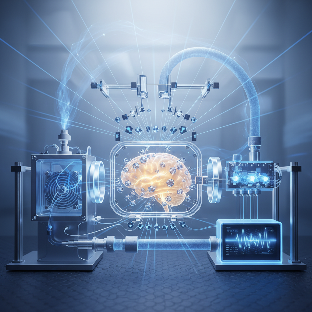
NV sensors placed within 5–20 nm of the source can transduce local magnetic spectral density into measurable changes in NV T1 and T2, and pulsed control lets us target specific noise bands (kHz–GHz). Dynamical decoupling acts both as a spectral filter and a tool: if biological coherence is present, selective sequences will amplify its signature versus broadband thermal noise.
Falsifiable predictions
- NV relaxometry will reveal narrowband peaks in the local magnetic spectral density at frequencies consistent with hyperfine/Zeemansplittings of candidate radical pairs; these peaks will persist at 295–310 K and collapse with selective chemical quenchers or targeted mutation of the photoreceptor.
- Application of tailored dynamical‑decoupling sequences (CPMG/XY8 families) will enhance NV‑detected signal amplitude and apparent coherence times only if the source dynamics are quantum‑coherent and spectrally narrow; purely classical broadband fluctuations will not show the same sequence dependence.
- Isotopic substitution (deuteration of nearby hydrogens) or controlled changes in static magnetic field strength (10–100 mT) will shift or damp the detected spectral features in ways predictable by radical‑pair Hamiltonians; null results set upper bounds on spin correlation amplitudes (e.g., magnetic field rms < 1 nT at 10 nm over 100 ns).
Experimental roadmap
1) Calibrate and benchmark: use test samples with known spin dynamics (single electron spins, organic radicals) to validate NV sensitivity (T1/T2, spectral reconstruction, spatial resolution). Use both ensemble and single NVs; employ scanning NV (AFM) for mapping.
2) Minimal model systems: isolated photoreceptor proteins (cryptochrome, flavin radical pairs) in controlled buffers on functionalized diamond surfaces with shallow NVs (<10 nm). Perform photoactivation and measure time‑resolved relaxometry and pulsed spectral scans.
3) Reduced complexity assemblies: photosynthetic complexes, reconstituted filamentous proteins (microtubules), and purified paramagnetic protein clusters to probe mesoscale signatures.
4) Cell and tissue tests: nanodiamond internalization into cultured cells expressing target proteins; or diamond chip substrates with adherent living cells/tissue slices. Compare live versus fixed, and temperature series.
5) Cross‑validation: correlate NV signals with ultrafast optical spectroscopy, EPR (when feasible), and functional readouts (behavioral assays for magnetoreception models).
Controls and potential pitfalls
Controls: heat‑killed cells, chemical radical scavengers, genetic knockouts/mutations of candidate proteins, isotopic substitution (D versus H), and external RF fields to intentionally disrupt hypothesized coherences. Use surface passivation and reference diamonds without shallow NVs to subtract diamond surface noise.
Pitfalls: spurious surface spin noise or magnetic contaminants may mask weak biological signals; proximity constraints limit sensitivity (signal decays ~1/r^3 for dipolar fields); motional averaging in liquids can wash out signatures; biological complexity can produce classical correlated noise mimicking quantum features. Mitigation includes optimized diamond surface chemistry, cryo‑control of select experiments (to validate temperature scaling), spectral tomography with multiple pulse sequences, and orthogonal measurement modalities (optical/EPR).
Implications
Positive detection of room‑temperature, functionally relevant quantum coherence would reframe mechanistic models in sensory biology, energetic transport, and biomolecular design — suggesting biology can engineer environments to preserve quantum resources. Negative but quantitatively stringent bounds will constrain radical‑pair and excitonic hypotheses and focus theory on classical or fast‑decohering mechanisms. Technologically, insights could inspire biomimetic strategies for ambient‑temperature quantum devices.
Summary: NV‑center quantum probes provide a concrete, tunable, and falsifiable toolkit to test whether warm biological systems exploit coherent quantum dynamics. The experiments outlined are technically demanding but within reach of current quantum sensing and molecular biology capabilities; they will yield definitive evidence or tight limits within a tractable experimental program.