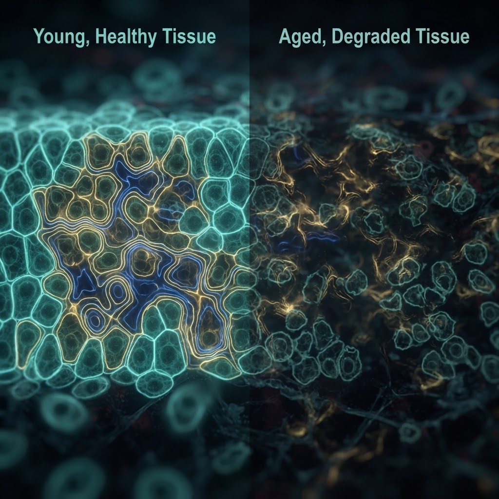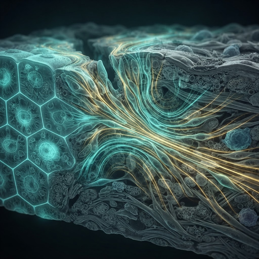
When skin is cut, the injury does more than break a physical barrier. It also disrupts the tissue’s electrical architecture. Intact epithelia maintain voltage differences through ion pumps, channels, and tight cell-cell junctions. At a wound, that circuit is suddenly shorted: current flows outward through the damaged region, producing an electric field at the wound edge.
These “electric wounds” are not merely a by-product of injury. A substantial body of work shows that endogenous voltage gradients can influence how cells orient, migrate, divide, and differentiate. The field is typically weak—on the order of tens to hundreds of millivolts per millimeter—but cells can detect it through voltage-sensitive signaling, membrane polarization, ion flux, and interactions with the cytoskeleton.
A directional cue in a crowded repair program
Wound healing is usually described as a sequence: clotting, inflammation, tissue formation, and remodeling. Bioelectricity cuts across all four stages. Immediately after epithelial damage, the electric field provides a directional cue that can help guide keratinocytes and other repair cells toward the wound. This directed migration, or galvanotaxis, is not simply faster movement. Cells can polarize their front and rear in response to the field, reorganizing actin networks and signaling molecules that control motility.
Ion channels and pumps are central to the process. Transporters that move sodium, potassium, chloride, and calcium across membranes establish local voltage differences and alter intracellular signaling. Calcium, in particular, can act as a rapid messenger linking electrical changes to gene expression, adhesion, and cytoskeletal remodeling. The result is a feedback system: injured cells change ionic flows, those flows reshape the field, and the field influences the behavior of neighboring cells.
From observation to intervention
Recent progress has come from combining live imaging, computational modeling, organoid systems, and tools that manipulate ion transport with increasing precision. Researchers can map voltage landscapes around wounds, suppress specific channels, or apply externally generated fields that mimic aspects of the endogenous signal. In laboratory models, controlled electrical stimulation has sometimes accelerated closure, redirected migration, or improved the organization of regenerating tissue.
The attraction is obvious for regenerative medicine. Chronic wounds—including diabetic ulcers—often involve impaired vascularization, inflammation, epithelial migration, and electrical signaling. Devices or biomaterials that restore a more favorable electrical environment could complement conventional approaches. Conductive hydrogels, electrode systems, and electrically responsive scaffolds are being investigated as ways to deliver spatially and temporally controlled cues rather than relying only on drugs or growth factors.
The limits of the “electric blueprint”
Yet voltage gradients are not a master switch for regeneration. Their effects depend on tissue type, developmental state, extracellular matrix, immune context, and the precise waveform and direction of stimulation. A field that helps a sheet of epithelial cells migrate may have different or unwanted consequences in a complex organ. Moreover, many experiments remain in cell culture or animal models, where geometry and tissue composition differ from human wounds.
The active debate is therefore shifting from whether bioelectricity matters to how it can be measured and controlled safely in living tissue. Researchers still need better maps of voltage dynamics over time, clearer separation of electrical effects from accompanying chemical signals, and clinical studies that identify which patients and wound types are most likely to benefit.
The emerging picture is nevertheless consequential. Wound healing is not governed only by chemical gradients and mechanical forces. It also unfolds within an electrical landscape generated by living cells. Understanding that landscape could make regeneration less a matter of waiting for tissue to repair itself—and more a matter of tuning the signals that tell it where, when, and how to grow.