Biophotons are back in the lab, and under sharper scrutiny
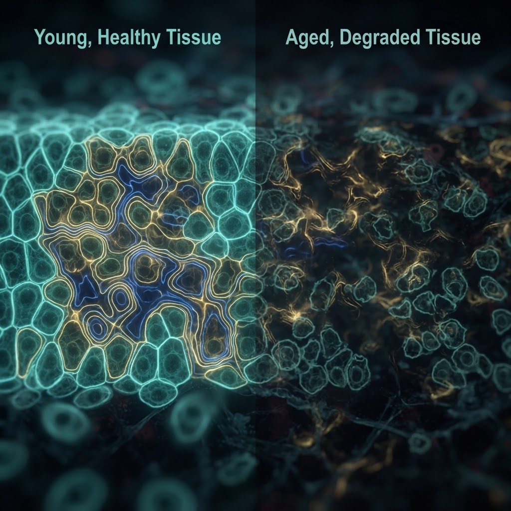
For five decades the idea that living cells emit and might use light to communicate sat at the margins of mainstream biology. That boundary is shifting. Improvements in low-noise photodetectors, time-resolved counting and spectral analysis have turned qualitative curiosities into quantitative datasets: cells, tissues and whole organisms produce measurable ultraweak photon emission (UPE), the intensity and spectrum of which track metabolic state and oxidative chemistry.
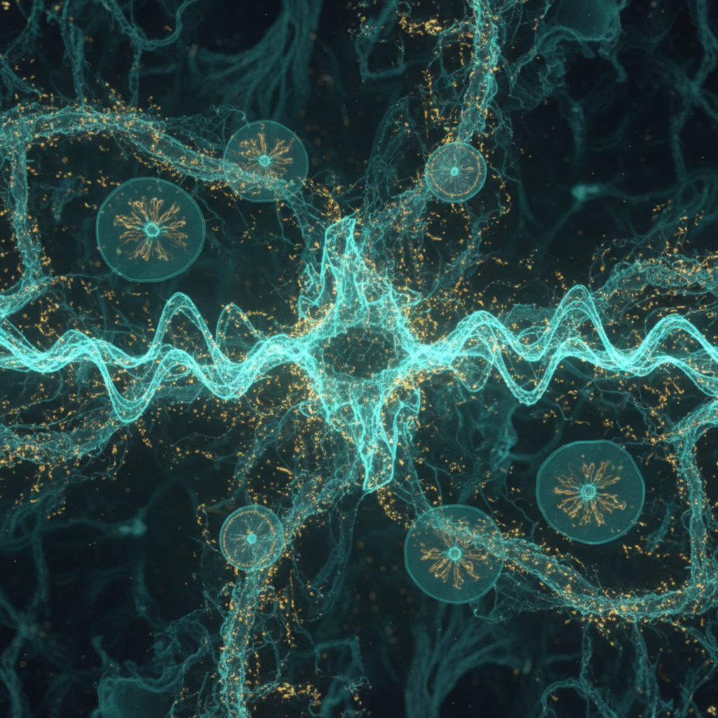
What has changed experimentally? Three converging trends. First, single-photon–sensitive instruments now permit reproducible detection of spontaneous emission from small cell populations and even neuronal preparations. Second, careful spectral and temporal correlation work shows that UPE correlates with mitochondrial activity and reactive oxygen species production—linking emission to canonical biochemistry rather than mystical "bioenergy." Third, new optical-modeling and microscopy studies have proposed plausible routes for photons to be guided inside biological structures such as myelinated axons or intracellular filaments, reducing the previous objection that tissue scattering would scatter any signal to oblivion.
Evidence and the cautious interpretation
Key experimental threads are clear but limited in scope. Observational studies repeatedly find that:
- UPE intensity rises with metabolic stimulation and oxidative stress and falls with antioxidants.
- Photon emission has spectral features—often in the visible to near-infrared—that reflect known chromophores and reaction intermediates.
- In vitro preparations can exhibit photon-mediated coupling effects under controlled geometries, suggesting photons can influence nearby cells under ideal conditions.
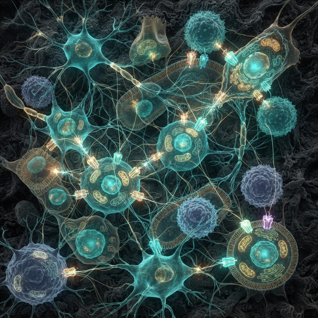
These robust associations support the proposition that biophotons are real, biologically linked phenomena. But the more extraordinary claim—that cells use photons as a controlled, information-rich intercellular signaling modality—remains unresolved. Demonstrating function requires not only emission, but a sender/receiver architecture, encoding, transmission fidelity in vivo, and receptor mechanisms sensitive to single-photon–level signals.
Why the question matters
If validated, photonic signaling would expand the repertoire of cellular communication. Light-based channels could operate on micro- to millisecond timescales and across distances where electrochemical signaling is slow or impractical. Practical implications include novel biomarkers of oxidative pathology (since UPE reflects redox state), new noninvasive diagnostics, and engineered bio-optical interfaces that exploit endogenous emission.
Points of contention and next steps
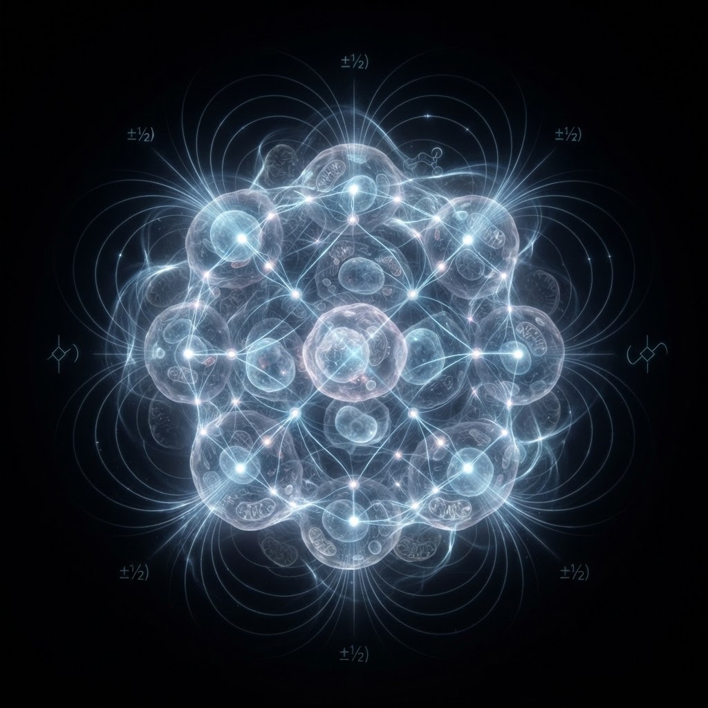
Healthy skepticism persists for good reasons. Claims of long-range coherence or quantum processing linked to biophotons outpace the data. Likewise, the identity of putative biological photoreceptors—opsin-like proteins, porphyrin/chromophore complexes or even structural waveguides—remains speculative. The field needs experiments that combine:
- in vivo, cell-type–specific photon recording synchronized to physiological events,
- loss- and gain-of-function tests for candidate photoreceptors, and
- manipulations that alter UPE independently of classical signaling pathways while testing downstream functional consequences.
Practically, initiatives that integrate single-photon detection with targeted optogenetic reporters, improved spectral fingerprinting and computational models of tissue optics will define whether biophotons are epiphenomena or elements of a light-based language in biology.
For now, the glow is real; whether it is gossip, noise, or conversation is the central question—and one the community is finally equipped to answer.