Why the periphery matters
Most popular explanations of circadian biology point to the suprachiasmatic nucleus (SCN) in the hypothalamus, the so-called master clock. That framing is useful but misleading: almost every metabolic tissue — liver, adipose, skeletal muscle, pancreas — contains autonomous molecular clocks that keep local time and control tissue-specific daily programs. These peripheral clocks do more than echo the SCN. They set the tempo for glucose handling, lipid synthesis, mitochondrial efficiency and hormone secretion.
How peripheral clocks regulate metabolism
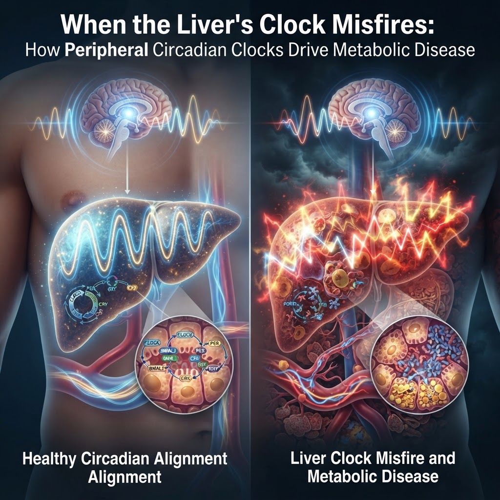
At the molecular level, clock proteins (CLOCK, BMAL1, PER, CRY, REV-ERB) form transcriptional-translational feedback loops that rhythmically open and close chromatin at genes involved in metabolism. The result is a daily choreography: enzymes rise when nutrients arrive, mitochondrial function peaks when energy demand is highest, and lipogenesis is scheduled away from fasting periods. Post-translational regulators — NAD+-dependent sirtuins, AMPK — tie cellular energy state into clock timing, creating bidirectional crosstalk between metabolism and timekeeping.
Evidence it goes wrong
Genetic and behavioural experiments make the case that peripheral-clock disruption is causal for metabolic disease. Mice with mutations in core clock genes develop obesity, hyperglycaemia and dyslipidaemia, linking clock circuitry to whole-body energy balance. More tellingly, uncoupling peripheral rhythms from the central clock — for example by feeding at the "wrong" time — is sufficient to produce fat gain and insulin resistance even without extra calories.
- Clock gene mutants show spontaneous metabolic syndrome, demonstrating that intact molecular oscillators are required for normal energy homeostasis.
- Time-restricted feeding in mice prevents weight gain and metabolic dysfunction on an obesogenic diet, without reducing calories — a potent demonstration that timing alone resets peripheral programs.
- Shift work and circadian misalignment in humans are epidemiologically associated with increased diabetes and cardiovascular risk, consistent with the effects seen in animal models.
Why peripheral clocks are an overlooked therapeutic target
The striking aspect is how local and context-dependent these clocks are. Liver clock disruption mainly derails glucose and lipoprotein rhythms; adipose clocks affect lipid storage and adipokines; pancreatic clocks tune insulin release. That modularity is an opportunity: therapies that restore synchrony — behavioral (timed feeding, light exposure), pharmacologic (REV-ERB agonists and other clock modulators) or even tissue-targeted interventions — could correct metabolic dysfunction without blunt systemic effects.
Complications and a cautionary note
It’s not simple. Peripheral clocks are redundant and plastic. Different tissues respond to different zeitgebers (feeding cues for liver and gut; activity and hormones for muscle and fat). A one-size-fits-all timing prescription is unlikely to work. Humans vary in chronotype, occupation and co-morbidities; what helps one person may harm another. Moreover, the extent to which peripheral misalignment is a primary cause versus a downstream marker of metabolic disease in humans remains incompletely resolved.
Hidden in plain sight: peripheral clocks are not a niche curiosity. They are local governors of metabolic flux, and when their rhythms slip out of sync with behaviour and the central clock, disease follows. The next decade should shift from asking whether circadian biology matters to asking how we safely and precisely re-synchronize tissue clocks in patients.