Neurons are built to last. Many survive for decades without dividing, extending vast networks of axons and synapses while continually manufacturing, transporting, and replacing their molecular parts. Their longevity is also their vulnerability. A damaged protein or worn-out mitochondrion cannot simply be diluted through cell division. It must be repaired, isolated, or destroyed. One of the principal systems responsible is autophagy—literally, “self-eating.”
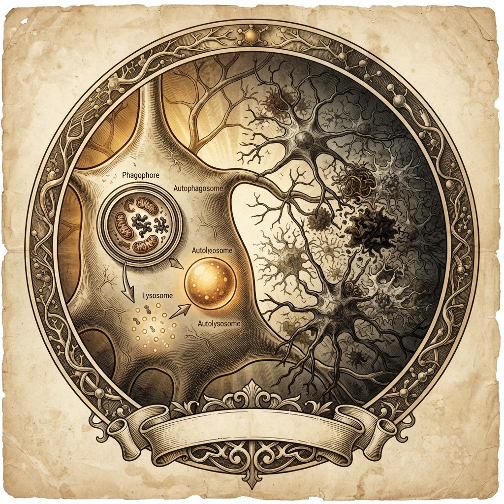
The phrase sounds destructive, but autophagy is better understood as cellular renewal. It encloses selected material in a double-membraned compartment, the autophagosome, which then fuses with a lysosome. Inside that acidic digestive organelle, proteins, lipids, and organelles are broken down and their components returned to the cell’s metabolic economy. In a neuron, this is not optional housekeeping. It is a continuous logistics operation extending from the cell body to distant nerve terminals.
From starvation to a Nobel-winning pathway
The modern story began in the 1950s, when Belgian cytologist Christian de Duve identified lysosomes and recognized them as intracellular digestion chambers. Electron microscopy soon revealed structures that appeared to engulf portions of the cytoplasm. De Duve introduced the term autophagy in 1963, initially describing a phenomenon associated with starvation and the cell’s effort to recycle its own resources.
For decades, the process remained important but technically elusive. Its molecular logic became clearer through the work of Japanese geneticist Yoshinori Ohsumi. In the 1990s, using baker’s yeast, Ohsumi identified genes required for autophagy and showed that a pathway first observed morphologically could be dissected genetically. The discovery established that autophagy is not a vague emergency response but a conserved, regulated program. Ohsumi received the 2016 Nobel Prize in Physiology or Medicine.
That history changed neuroscience. Researchers began to ask whether autophagy might explain why neurons accumulate the very materials they should be clearing: amyloid-beta and tau in Alzheimer’s disease, alpha-synuclein in Parkinson’s disease, huntingtin fragments in Huntington’s disease, and misfolded proteins associated with amyotrophic lateral sclerosis.
Why neurons are especially dependent on it
Autophagy has an unusual geography in the nervous system. Autophagosomes often form at the ends of axons, where damaged components arise far from the cell body. They must then travel along microtubules, mature, and meet lysosomes. A failure at any point—formation, cargo selection, transport, fusion, or degradation—can leave neurons crowded with partially processed material.
This makes autophagy an attractive therapeutic target. In principle, drugs could increase autophagosome formation, improve lysosomal function, enhance the removal of particular toxic proteins, or restore transport along axons. The mammalian target of rapamycin, or mTOR, is a major nutrient-sensing regulator that generally suppresses autophagy when resources are abundant. Rapamycin and related compounds can stimulate autophagy by inhibiting mTOR, while other experimental agents act on lysosomal acidity, transcriptional regulators, or selective cargo receptors.
Yet “more autophagy” is not automatically better. Autophagy is a sequence, not a single switch. Increasing the production of autophagosomes without improving their clearance can worsen congestion. In some disease states, lysosomes are already overloaded or damaged; pushing more cargo into a broken disposal system may be counterproductive. The therapeutic goal is therefore not indiscriminate activation, but restoration of flux—the successful movement of material from capture to degradation and recycling.
Promise, evidence, and caution
Animal studies have supplied compelling clues. Genetic enhancement of autophagy can reduce toxic protein accumulation and, in some models, improve neuronal survival. Conversely, disrupting essential autophagy genes in the nervous system causes progressive neurodegeneration. These findings make the pathway biologically credible, but they do not guarantee a useful medicine. Mouse models often reproduce protein aggregates more readily than they reproduce the decades-long evolution, cellular diversity, and clinical complexity of human disease.
Human trials have consequently produced a mixed picture. Rapamycin-like drugs face challenges involving immune suppression, metabolic effects, and dosing. Autophagy-modulating agents such as trehalose, lithium, and various lysosomal or mTOR-directed compounds have generated intriguing laboratory results, but robust clinical benefit remains unproven for most neurodegenerative conditions. Measuring autophagy in living human brains is itself difficult, making it hard to know whether a treatment reaches the right cells and changes the pathway in the intended direction.
The next chapter: precision cleanup
The field is moving toward selectivity. Rather than broadly stimulating cellular self-digestion, researchers are developing approaches that target particular cargo or organelles: enhancing mitophagy to remove defective mitochondria, correcting lysosomal storage defects, or designing “autophagy-tethering” molecules that bring disease proteins to the degradation machinery. Gene therapies may eventually tune pathway components in defined neuronal populations, although delivery and long-term safety remain formidable obstacles.
Autophagy matters today because it reframes neurodegeneration. These diseases are not only problems of toxic protein production; they are also failures of traffic, quality control, and recycling. The foundational insight from de Duve, extended through Ohsumi’s genetic work, has become a therapeutic proposition: neurons may be protected not merely by blocking what harms them, but by repairing the systems that clear harm away. The open question is whether medicine can make that intervention precise enough—enhancing renewal without provoking damage—to turn cellular housekeeping into durable neurological recovery.