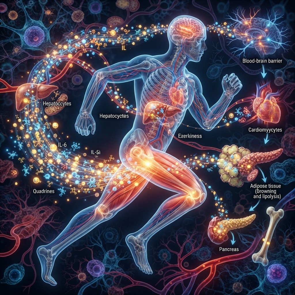
Exercise is not only a local stress imposed on skeletal muscle. It is a whole-body signaling event. As muscles contract, and as organs sense changing energy demand, temperature, oxygen availability, and mechanical load, cells release molecules that alter the behavior of distant tissues. This emerging language of exercise-induced communication is increasingly known as exerkine signaling.
The term “exerkine” covers a diverse group of exercise-responsive factors, including cytokines, peptides, metabolites, lipids, nucleic acids, and extracellular vesicles. Some are released into the circulation; others act close to the tissue that produces them. Together, they help coordinate fuel use, inflammation, vascular function, tissue repair, and adaptation to repeated training.
Muscle as an endocrine organ
The best-established example is interleukin-6 (IL-6). Contracting muscle can release IL-6, particularly during prolonged or strenuous exercise. In the acute setting, IL-6 helps mobilize energy substrates and can influence glucose and lipid metabolism in the liver and adipose tissue. Its effects are context-dependent: a brief exercise pulse is biologically different from persistently elevated IL-6 associated with chronic inflammation.
Other muscle-derived signals include irisin, generated from the membrane protein FNDC5, and myostatin, a regulator of muscle growth. Irisin has been linked in experimental models to changes in adipose tissue and bone biology, while myostatin restrains muscle mass. Yet the clinical picture remains more complicated than early headlines suggested. Human concentrations, assay reliability, tissue sources, and the size of effects are still debated for several proposed myokines.
A network, not a single “exercise hormone”
Exercise-induced communication also runs in the opposite direction. The liver releases hepatokines that influence muscle metabolism; adipose tissue produces adipokines and lipid mediators; bone releases factors such as osteocalcin; and the brain and vascular endothelium respond to, and may contribute to, circulating signals. Exercise can also alter extracellular vesicles carrying proteins, lipids, and regulatory RNA between tissues.
Small metabolites may be just as important as classical hormones. Lactate, once treated mainly as a waste product, can serve as a fuel and signaling molecule. During exercise it can affect gene expression, vascular responses, and neuronal function. Succinate and other intermediates of cellular metabolism can also act as signals when their concentrations shift. These molecules provide a rapid biochemical connection between energy production and longer-term adaptation.
Why the field is moving now
Recent advances in single-cell sequencing, spatial biology, metabolomics, and high-sensitivity proteomics are allowing researchers to identify which cells produce a signal, which tissues receive it, and how responses vary with exercise intensity, duration, sex, age, training status, and disease. This matters because exercise is not one intervention. A sprint, a long run, resistance training, and rehabilitation produce overlapping but distinct molecular signatures.
The promise is practical. Exerkines could help explain why physical activity improves insulin sensitivity, cardiovascular health, brain function, and immune regulation. They may also provide biomarkers for whether a training program is biologically effective, or inspire therapies that mimic selected benefits of exercise for people unable to exercise adequately.
But the field faces a critical test: association is not proof of mediation. A molecule that rises after exercise may be a marker of stress rather than the cause of adaptation. Many findings from rodents have not translated cleanly to humans, and commercial assays can produce inconsistent measurements. Dose, timing, tissue specificity, and combinations of signals may matter more than any single factor.
The current view is therefore less a hunt for one “exercise pill” than an attempt to decode a dynamic conversation. Exerkine research is beginning to map that conversation—one in which organs continually report their state, negotiate resources, and reshape one another in response to movement.